Download
1 / 13
150 likes | 370 Views
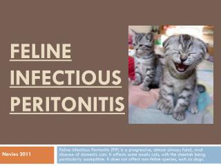
F eline I nfectious P eritonitis. Accession #137373 Christina Copple, DVM. “Fiver”, 5mth old, Male, Singapura. Clinical Complaint: Chronic inappetance with acute onset anorexia, vomited once? PE abnormalities: Decreased to absent lung sounds Prominent LNs on palpation

E N D
Feline Infectious Peritonitis Accession #137373 Christina Copple, DVM
“Fiver”, 5mth old, Male, Singapura • Clinical Complaint: Chronic inappetance with acute onset anorexia, vomited once? • PE abnormalities: • Decreased to absent lung sounds • Prominent LNs on palpation • Abdomen distended, palpable fluid wave • Fast US in ER: moderate peritoneal & pleural effusion
“He’s Got Fluid” • Thoracocentesis & Abdominocentesis • yellow, viscous fluid • TP = 4.8 • Modified transudate with disproportionate increase in protein • + Rivalta precipitates = more consistent with exudate
Feline Infectious Peritonitis • Mutated Feline Enteric Coronavirus • Risk Factors: • Mutlicat household or cattery • Purebred - cattery • Sexually intact • Young to middle aged (<5yrs) • Higher incidence (75%) in males • 2 Forms: • Effusive • Noneffusive
Effusive Form ----- Fiver • Immune-mediated vasculitis • Activated macrophages which contain actively replicating FIP virus • Loss of protein-rich fluid: peritoneal effusion, pleural effusion or pericardial effusion • Subcapsular space of kidneys • Effusion – high-protein, moderately cellular == modified transudate
Noneffusive Form • Pyogranulomatous or Granulomatous Inflammation • Multiple organs affected • Eyes • Brain • Kidney • Omentum • Focal intestinal lesions
Abdominal Ultrasonographic Findings Associated with Feline Infectious Peritonitis: A retrospective Review of 16 cases • 13 had necropsy, remaining 3 were combo of histo, cyto, and clinpath findings highly suggestive of FIP • Normal US does not exclude possibility of FIP • No US findings were specific or sensitive for hepatic or splenic changes associated with FIP • Liver: normal in 11 (69%), diffusely hypoechoic in 3 (19%), focally hyperechoic in 1 (0.6%) and focally hypoechoic in 1 (0.6%) • Spleen: normal in 14 (88%), hypoechoic in 2 (1.2%) JAAHA 2010;46:152-160
Abdominal Ultrasonographic Findings Associated with Feline Infectious Peritonitis: A retrospective Review of 16 cases • With appropriate clinical signs and a combination of the following findings, the index of suspicion for FIP should increase • Renomegaly • Irregular renal contour & hypoechoic subcapsular echogenicity • Abdominal lymphadenopathy • Peritoneal or retroperitoneal effusion • Diffuse changes within intestines JAAHA 2010;46:152-160
Figure 1—Transverse ultrasound image of a left kidney showing irregular renal margins, hypoechoic subcapsular infiltration (white arrows), and mottled echogenicity. Figure 2—Ultrasound image of a right kidney demonstrating mottled renal echogenicity, hypoechoic subcapsular infiltration (white arrows), and decreased corticomedullary definition. Both kidneys were subjectively enlarged. JAAHA 2010;46:152-160
Abdominal Ultrasonographic Findings Associated with Feline Infectious Peritonitis: A retrospective Review of 16 cases • Pyogranulomatous or granulomatous hepatitis, nephritis, or pleuritis • Pleural & peritoneal effusion • Pyogranulomatousorchitis • Pyogranulomatous enteritis • Diffuse intestinal involvement more than solitary lesions • 3 (19%) intestinal thickening • More often small intestine vs large intestine • 1 - diffuse small intestinal thickening • 1 - duodenal thickening • 1 – colonic thickening with loss of layering JAAHA 2010;46:152-160