Download
1 / 63
630 likes | 676 Views
Bones Quiz - What bones comprise the wrist? Joints Quiz - What joints comprise the wrist?. Wrist Anatomy. Proximal Row Where can you palpate these? Scaphoid Lunate Triquetrum Pisiform Radiocarpal joint Ulnocarpal joint Intercarpal joints. Distal Row Where can you palpate these?

E N D
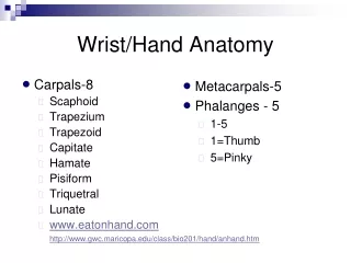
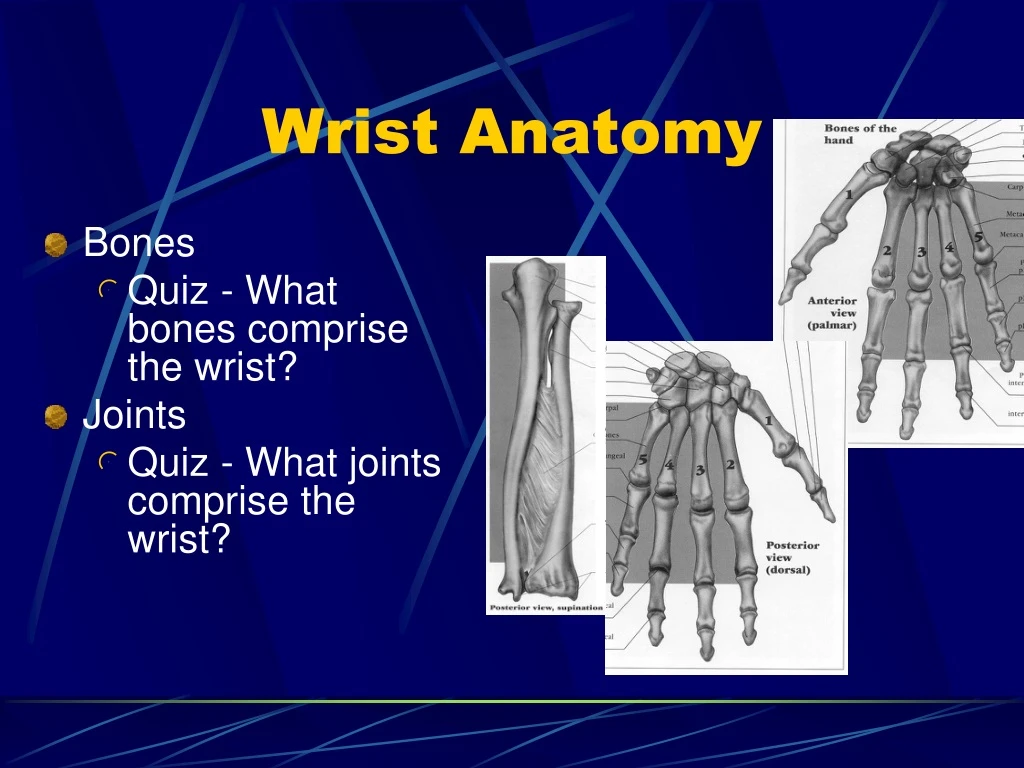
Bones Quiz - What bones comprise the wrist? Joints Quiz - What joints comprise the wrist? Wrist Anatomy
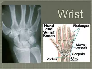
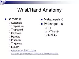
Proximal Row Where can you palpate these? Scaphoid Lunate Triquetrum Pisiform Radiocarpal joint Ulnocarpal joint Intercarpal joints Distal Row Where can you palpate these? Trapezium Trapezoid Capitate Hamate Intercarpal joints Carpometacarpal joints (related to hand) Carpal Bones and Articulations
Articulations and ROM • Distal Radioulnar joint • Supination and Pronation – 80-90o • Ulna moves posteriorly and laterally with pronation • Radiocarpal joint (and Ulnocarpal joint) • Flexion (80-90o) and Extension (75-85o) • Radial (20o) and Ulnar (35o) Deviation • Intercarpal joints • Gliding
Ligaments Covered by a fibrous capsule Radial and ulnar collateral limit ulnar and radial deviation; collectively limits flexion and extension Intercarpal and Carpometacarpal Soft tissue of Wrist
Ligaments Dorsal – limits flexion Dorsal Radiocarpal Palmar - limit extension Transverse carpal ligament Palmar radiocarpal Multiple divisions Palmar ulnocarpal ligament Multiple divisions Soft tissue of Wrist
Cartilage Triangular Fibrocartilage Complex – TFCC “Meniscus” between ulna and triquetrum Ulnar collateral ligament and palmar ulnocarpal ligaments have attachments Compressed with Pronation and Extension Compressed with Ulnar deviation Soft tissue of Wrist
Extensor muscles Extensor Retinaculum What’s its function? Muscles innervated by radial nerve What are they? Name them… Flexor Muscles Flexor retinaculum (aka transverse carpal ligament) Two compartments Superficial Deep Innervated by median and ulnar nerve Muscle Tissue of Wrist
EXTENSORS FLEXORS
Wrist and Hand Anatomy • Nerves/Vessels • Radial & ulnar artery and veins • Radial, ulnar, & median nerves • Carpal Tunnel - • Flexor Tendons - 9 • Median Nerve
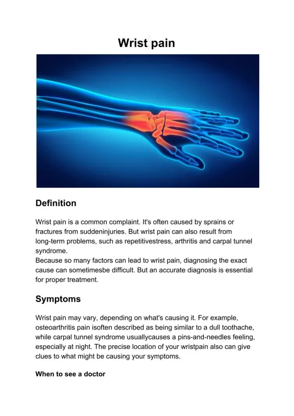
Wrist Injuries • Strains • Onset usually acute – FOOSH or Overexertion • S/S: Active ROM limited • Wrist Ganglion • Herniation of the joint capsule or synovial sheath of a tendon. Tx: Bible Therapy
Wrist Injuries • deQuervain’s Disease - thumb/wrist • stenosing tenosynovitis of the extensor pollicis brevis and abductor pollicis longus. • S/S: crepitation, tenderness, strength loss. • Special Test: = Finkelstein’s test • Tx: RICE, NSAIDs
Wrist Injuries • Sprains • Onset is usually acute – FOOSH or overexertion • Often diagnosed when other injuries are ruled out • Both active and passive ROM are effected • S/S: Laxity, pain, swelling, limited ROM • Pain is usually with overstretching • Special Tests: Varus/Valgus, Carpal Glide • PRICE, Rehabilitation, Taping for prevention
Wrist Injuries • Triangular Fibrocartilage Injuries - TFCC • Onset is usually acute • MOI: Forced hyperextension of wrist with loading • S/S: Pain with pronation/extension and/or ulnar deviation; Pain with loading; Point tenderness; Swelling; Altered joint mechanics • Special Test: Valgus test elicits pain but no laxity and Varus test compresses and causes pain • Immobilization and Surgery are often necessary
Neural Injuries • Carpal Tunnel Syndrome • Compression of median nerve • Fibrosis of the synovium of flexor tendons secondary to tenosynovitis • MOI: Insidious onset with repetitive wrist movement (and finger movement); Acute onset with trauma; Progressive degeneration • S/S: numbness palmar thumb, index, middle fingers, dull ache, weak finger flexion (grip). May worsen with sleep. • Poor posture may predispose. • Special Tests: Tinel’s sign and Phalen’s • Tx: Conservative (PRICE, NSAIDs) and Surgical
Neural Injuries • Biker’s Palsy • Ulnar nerve compression • Ulnar nerve passes through tunnel of Guyon between pisiform and hamate. • MOI: repetitive jarring or pressure, repetitive flx/ext/ulnar deviation • Tx: Padding (Gloves), Ice, NSAIDs • Drop Wrist Syndrome • Radial nerve compression at elbow • Inability to extend wrist and fingers
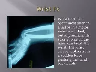
Wrist Injuries • Wrist Fractures • Distal Radius/Ulna and Forearm Fractures • Onset is acute • MOI: Hyperextension or hyperflexion combined with rotatory motion – FOOSH • S/S: Deformity felt and observed; Crepitus • Evaluated Neurovascular status • Tx: Splint, Ice, Referral
Wrist Injuries • Wrist Fractures • Distal Radius/Ulna • Colles’ Fracture • MOI: hyperextension-fall on outstretched • S/S: “silver fork deformity” - radius & ulna posteriorly • Smith’s Fracture (Reverse Colles) • MOI: hyperflexed • S/S: “garden spade deformity” - radius & ulna anteriorly
Wrist Fractures Scaphoid - most common carpal MOI: fall on outstretched hand S/S: wrist aches, pain in anatomical snuff box, painful handshake or with overpressure Tx: Splint, Referral, Ice Plain X-rays may not be enough Immobilization (long and/or short) – 12 weeks Risk: aseptic necrosis and non-union fractures Preiser’s Disease Surgery may be necessary Wrist Injuries
Wrist Dislocations Radius or Ulna Lunate is very common MOI: force hyperextension Dorsal displacement = perilunate dislocation Palmar displacement (total rupture) = lunate dislocation S/S: Deformity, 3rd Knuckle is lower (Murphy’s sign), Paresthesia of middle finger, weak finger flexion Risk: Untreated or repeated trauma Kienbock’s Disease Decreased grip, pain with ulnar deviation, weak extension, pain with passive 3rd finger extension Immobilization – 6-8 weeks; Surgery may be necessary Wrist Injuries
Wrist Injury Prevention • Good technique! • But…these help
Hand and Fingers Pathology
4 1 3 2 Flexor tendon arrangement Lumbricals Dorsal Interossei Palmar Interossei
Extensor Hood, Long extensor tendon, and lateral bands Finger flexor tendons Unique finger Look at pulley system
Observation • Relaxed position of hand • Fingers slightly flexed • Relative shortness of finger flexors • Skin and Nail health • Discoloration, texture, hair patterns • Finger alignment • Tips of fingers should align with finger flexion • Hand abnormalities • Finger and metacarpal positioning • Muscle atrophy • Range of motion
Range of Motion • Carpometacarpal • Flexion (70-80o)/Extension • Abduction (70-80o)/Adduction • Opposition • Metacarpophalangeal • Flexion (85-105o)/Extension (20-35o) • Abduction/Adduction (20-25o) • Interphangeal joints • Thumb flexion (80-90o) • PIP flexion (110-120o) • DIP flexion (80-90o)
Palpation • Metacarpals and joints • Collateral ligaments of MCPs • Phalanges and joints • Collateral ligaments of PIPs and DIPs • Thenar compartment • muscles • Thenar webspace • muscles • Central compartment • Palmar fascia and muscles • Hypothenar compartment • muscles
Dupuytren’s Contracture Pathology • Tendon pathology • Trigger Finger/Thumb • Mallet Finger • Boutonniere Deformity • Jersey Finger • Dupuytren’s Contracture • Swan Neck Deformity • Joint pathology • Sprains • Bony pathology • Fractures • Dislocations Swan Neck Deformity
Tendon pathology • Trigger Finger or Thumb • Etiology • Repeated motion of fingers may cause irritation, producing tenosynovitis • Inflammation of tendon sheath (flexor tendons of wrist, fingers and thumb, abductor pollicis) • Thickening forming a nodule that does not slide easily • Signs and Symptoms • Resistance to re-extension, produces snapping that is palpable, audible and painful • Palpation produces pain and lump can be felt w/in tendon sheath • Management • Immobilization, rest, cryotherapy and NSAID’s • Ultrasound and ice are also beneficial • Injection
Tendon pathology • Mallet Finger (baseball or basketball finger) • Etiology • Caused by a blow that contacts tip of finger avulsing extensor tendon from insertion • Avulses extensor digitorum at distal phalanx • Signs and Symptoms • Unable to extend distal end of finger (carrying at 30 degree angle) • Point tenderness at sight of injury • X-ray shows avulsed bone on dorsal proximal distal phalanx • Management • RICE and splinting in hyperextension for 6-8 weeks
Tendon pathology • Boutonniere Deformity • Etiology • Rupture of extensor tendon dorsalto the middle phalanx – bone passes through central slip • Forces DIP joint into extension and PIP into flexion • Signs and Symptoms • Severe pain, obvious deformity and inability to extend DIP joint • Swelling, point tenderness • Management • Cold application, followed by splinting in PIP extension and DIP flexion • Splinting must be continued for 5-8 weeks
Tendon pathology • Jersey Finger • Etiology • Rupture of flexor digitorum profundus tendonfrom insertion on distal phalanx • Often occurs w/ ring finger when athlete tries to grab a jersey • Signs and Symptoms • DIP can not be flexed, finger remains extended • Pain and point tenderness over distal phalanx • Management • Must be surgically repaired • Rehab requires 12 weeks and there is often poor gliding of tendon, w/ possibility of re-rupture
Dupuytren’s Contracture Tendon pathology • Dupuytren’s Contracture • Etiology • Nodules develop in palmer aponeurosis, limiting finger extension - ultimately causing flexion deformity • Signs and Symptoms • Often develops in 4th or 5th finger (flexion deformity) • Management • Tissue nodules must be removed as they can ultimately interfere w/ normal hand function
Tendon pathology • Swan Neck Deformity Etiology • Distal tear of volar plate or finger trauma may cause Swan Neck deformity • Flexed MCP, extended PIP, and flexed DIP • Signs and Symptoms • Pain, swelling w/ varying degrees of hyperextension • Tenderness over volar plate of PIP • Indication of volar plate tear = passive hyperextension • Management • RICE and analgesics • Splint in PIP 20-30 degrees of flexion/DIP extension for 3 weeks; followed by buddy taping
Joint pathology • Sprains Phalanges • Etiology • Phalanges are prone to sprains caused by direct blows or twisting • Signs and Symptoms • Recognition primarily occurs through history • Sprain symptoms - pain, severe swelling and hemorrhaging
Joint pathology • Gamekeeper’s Thumb • Etiology • Sprain of UCL of MCP joint of the thumb • Mechanism is forceful abduction of proximal phalanx occasionally combined w/ hyperextension • Signs and Symptoms • Pain over UCL in addition to weak and painful pinch • Management • Immediate follow-up must occur • If instability exists, athlete should be referred to orthopedist • If stable, X-ray should be performed to rule out fracture • Thumb splint should be applied for protection for 3 weeks or until pain free • Splint should extend from wrist to end of thumb in neutral position • Thumb spica should be used following splinting for support
Joint pathology • Sprains of Interphalangeal Joints of Fingers • Etiology • Can include collateral ligament, volar plate, extensor slip tears • Occurs w/ axial loading or valgus/varus stresses • Signs and Symptoms • Pain, swelling, point tenderness, instability • Valgus and varus tests may be possible • Management • RICE, X-ray examination and possible splinting • Splint at 30-40 degrees of flexion for 10 days • If sprain is to the DIP, splinting for a few days in full extension may assist healing process • Taping can be used for support
PIP Dorsal Dislocation Etiology Hyperextension that disrupts volar plate at middle phalanx Signs and Symptoms Pain and swelling over PIP Obvious deformity, disability and possible avulsion Management Treated w/ RICE, splinting and analgesics followed by reduction After reduction, finger is splinted at 20-30 degrees of flexion for 3 weeks -- followed by buddy taping PIP Palmar Dislocation Etiology Caused by twist while it is semiflexed Signs and Symptoms Pain and swelling over PIP; point tenderness over dorsal side Finger displays angular or rotational deformity Management Treat w/ RICE, splinting and analgesics followed by reduction Splint in full extension for 4-5 weeks after which it is protected for 6-8 weeks during activity Joint pathology
Joint pathology • MCP Dislocation • Etiology • Caused by twisting or shearing force • Signs and Symptoms • Pain, swelling and stiffness at MCP joint • Proximal phalanx is angulated at 60-90 degrees • Management • RICE, following reduction splinting in slight flexion (3 weeks) • Buddy taping following splinting • Therapy
Bony Pathology • Metacarpal Fracture • Etiology • Direct axial force or compressive force • Fractures of the 5th metacarpal = Boxer’s Fracture • Signs and Symptoms • Pain and swelling; possible angular or rotational deformity • Management • RICE, analgesics are given followed by X-ray examinations • Deformity is reduced, followed by splinting - 4 weeks of splinting after which therapy starts • Unstable fracture may need to be surgically pinned
Bony pathology • Bennett’s Fracture • Etiology • Occurs at carpometacarpal joint of the thumb as a result of an axial and abduction force to the thumb • Signs and Symptoms • CMC may appeared to be deformed - X-ray will indicate fracture • Athlete will complain of pain and swelling over the base of the thumb • Management • Structurally unstable and must be referred to an orthopedic surgeon • Surgery and immobilization – season ending
Bony pathology • Distal Phalangeal Fracture • Etiology • Crushing force • Signs and Symptoms • Complaint of pain and swelling of distal phalanx • Subungual hematoma is often seen in this condition • Management • RICE and analgesics are given • Protective splint is applied as a means for pain relief • Subungual hematoma is drained
Bony pathology • Middle Phalangeal Fracture • Etiology • Occurs from direct trauma or twist • Signs and Symptoms • Pain and swelling w/ tenderness over middle phalanx • Possible deformity; X-ray will show bone displacement • Management • RICE and analgesics • No deformity - buddy tape w/ splint for activity • Deformity - immobilization for 3-4 weeks and a protective splint for an additional 9-10 weeks during activity
Bony pathology • Proximal Phalangeal Fracture • Etiology • May be spiral or angular • Signs and Symptoms • Complaint of pain, swelling, deformity • Inspection reveals varying degrees of deformity • Management • RICE and analgesics are given as needed • Fracture stability is maintained by immobilization of the wrist in slight extension, MCP in 70 degrees of flexion and buddy taping
Lacerations • Superficial location of tendons and nerves predisposes athletes to damage form shallow lacerations. • Any laceration to the fascia below the cutaneous layer should receive a referral • R/O trauma to tendons and nerves • Prevent infection • Suture to ensure minimal scarring
Finger Nail Pathology • Subungual Hematoma • MOI: finger caught between two surfaces • Presents with bleeding under nail bed • Draining – Drill or Cauterize • Paronychia • Infection around fingernail beds • S/S: Redness, pain, drainage • Warm soaks (Betadine), Antibiotic, Referral • Changes in normal appearance - indicative of a number of different diseases • Scaling or ridging = psoriasis • Ridging and poor development = hyperthyroidism • Clubbing and cyanosis = congenital heart disorders or chronic respiratory disease • Spooning or depression = chronic alcoholism or vitamin deficiency
Prevention of Hand Injuries • Protection • Gloves, Grips, Braces • Proper Technique • Sport and Ergonomics • Physical Conditioning • Reps and Sets for muscles of Hand • Theraputty, Wrist curls/extensions, Fist pumps