Download
1 / 77
790 likes | 830 Views
The Special Senses ( Hearing , Smell, Sight, Taste and Balance ). CLASS: I M.Sc., UNIT: 4 PREPARED BY: A. BENNO SUSAI. The Special Senses. Taste, smell, sight, hearing, and balance Special sensory receptors Localized – confined to the head region

E N D
The Special Senses(Hearing, Smell, Sight, TasteandBalance) CLASS: I M.Sc., UNIT: 4 PREPARED BY: A. BENNO SUSAI DEPARTMENT OF BIOCHEMISTRY, SJC, TRICHY
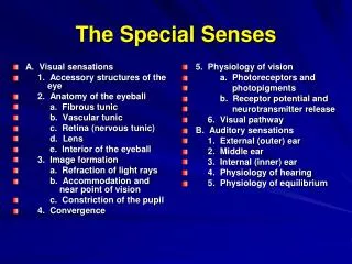
The Special Senses • Taste, smell, sight, hearing, and balance • Special sensory receptors • Localized – confined to the head region • Receptors are not free endings of sensory neurons • Special receptor cells DEPARTMENT OF BIOCHEMISTRY, SJC, TRICHY
The Chemical Senses: Taste and Smell • Taste – gustation • Smell – olfaction • Receptors – classified as chemoreceptors • Respond to chemicals DEPARTMENT OF BIOCHEMISTRY, SJC, TRICHY
Taste – Gustation • Taste receptors • Occur in taste buds • Most are found on the surface of the tongue • Located within tongue papillae DEPARTMENT OF BIOCHEMISTRY, SJC, TRICHY
Taste Buds • Collection of 50-100 epithelial cells • Contain three major cell types • Supporting cells • Gustatory cells • Basal cells • Contain long microvilli – extend through a taste pore DEPARTMENT OF BIOCHEMISTRY, SJC, TRICHY
Taste Buds DEPARTMENT OF BIOCHEMISTRY, SJC, TRICHY Figure 16.1a, b
Taste Sensation and the Gustatory Pathway • Four basic qualities of taste • Sweet, sour, salty, and bitter • A fifth taste – umami – “deliciousness” • No structural difference among taste buds DEPARTMENT OF BIOCHEMISTRY, SJC, TRICHY
Gustatory Pathway • Taste information reaches the cerebral cortex • Primarily through the facial (VII) and glossopharyngeal (IX) nerves • Some taste information through the vagus nerve (X) • Sensory neurons synapse in the medulla • Located in the solitary nucleus DEPARTMENT OF BIOCHEMISTRY, SJC, TRICHY
Gustatory Pathway from Taste Buds DEPARTMENT OF BIOCHEMISTRY, SJC, TRICHY Figure 16.2
Smell (Olfaction) • Receptors are part of the olfactory epithelium • Olfactory epithelium composed of: • Cell bodies of olfactory receptor cells • Supporting cells – columnar cells • Basal cells – form new olfactory receptor cells DEPARTMENT OF BIOCHEMISTRY, SJC, TRICHY
Smell (Olfaction) • Axons of olfactory epithelium • Gather into bundles – filaments of the olfactory nerve • Pass through the cribriform plate of the ethmoid bone • Attach to the olfactory bulbs DEPARTMENT OF BIOCHEMISTRY, SJC, TRICHY
Olfactory Receptors DEPARTMENT OF BIOCHEMISTRY, SJC, TRICHY Figure 16.3a, b
The Eye and Vision • Visual organ – the eye • 70% of all sensory receptors are in the eyes • 40% of the cerebral cortex is involved in processing visual information DEPARTMENT OF BIOCHEMISTRY, SJC, TRICHY
AccessoryStructures of the Eye • Lacrimal apparatus – keeps the surface of the eye moist • Lacrimal gland – produces lacrimal fluid • Lacrimal sac – fluid empties into nasal cavity DEPARTMENT OF BIOCHEMISTRY, SJC, TRICHY Figure 16.5b
The Fibrous Tunic • Most external layer of the eyeball • Composed of two regions of connective tissue • Sclera – posterior five-sixths of the tunic • White, opaque region • Provides shape and an anchor for eye muscles • Cornea – anterior one-sixth of the fibrous tunic • Limbus – junction between sclera and cornea • Scleral venous sinus – allows aqueous humor to drain DEPARTMENT OF BIOCHEMISTRY, SJC, TRICHY
Medial View of the Eye DEPARTMENT OF BIOCHEMISTRY, SJC, TRICHY Figure 16.7a
The Vascular Tunic • The middle coat of the eyeball • Composed of choroid, ciliary body, and iris • Choroid – vascular, darkly pigmented membrane • Forms posterior five-sixths of the vascular tunic • Brown color – from melanocytes • Prevents scattering of light rays within the eye • Choroid corresponds to the arachnoid and piamaters DEPARTMENT OF BIOCHEMISTRY, SJC, TRICHY
Posterior View of the Anterior Half of the Eye DEPARTMENT OF BIOCHEMISTRY, SJC, TRICHY Figure 16.9a
The Vascular Tunic • Ciliary body – thickened ring of tissue – encircles the lens • Composed of ciliary muscle • Ciliary processes – posterior surface of the ciliary body • Ciliary zonule (suspensory ligament) • Attached around entire circumference of the lens DEPARTMENT OF BIOCHEMISTRY, SJC, TRICHY
The Vascular Tunic DEPARTMENT OF BIOCHEMISTRY, SJC, TRICHY Figure 16.8
The Iris • Visible colored part of the eye • Attached to the ciliary body • Composed of smooth muscle • Pupil – the round, central opening • Sphincter pupillae muscle (constrictor or circular) • Dilator pupillae muscle (dilator or radial) • Act to vary the size of the pupil DEPARTMENT OF BIOCHEMISTRY, SJC, TRICHY
The Sensory Tunic (Retina) • Retina – the deepest tunic • Composed of two layers • Pigmented layer – single layer of melanocytes • Neural layer – sheet of nervous tissue • Contains three main types of neurons • Photoreceptor cells • Bipolar cells • Ganglion cells DEPARTMENT OF BIOCHEMISTRY, SJC, TRICHY
Microscopic Anatomy of the Retina Ganglion cells DEPARTMENT OF BIOCHEMISTRY, SJC, TRICHY Figure 16.10a
Photoreceptors • Two main types • Rod cells – more sensitive to light • Allow vision in dim light • Cone cells – operate best in bright light • Enable high-acuity, color vision • Considered neurons DEPARTMENT OF BIOCHEMISTRY, SJC, TRICHY
Photoreceptors DEPARTMENT OF BIOCHEMISTRY, SJC, TRICHY Figure 16.11
Rhodopsin – Visual purple Rhodopsin Bathorhodopsin Lumirhodopsin Metarhodopsin I Metarhodopsin II All – trans retinal All – trans retinol (Vitamin A) Scotopsin 11 – cis retinal Photoisomerization 11 – cis retinol DEPARTMENT OF BIOCHEMISTRY, SJC, TRICHY
Regional Specializations of the Retina • Macula lutea – contains mostly cones • Fovea centralis – contains only cones • Region of highest visual acuity • Optic disc – blind spot DEPARTMENT OF BIOCHEMISTRY, SJC, TRICHY
Blood Supply of the Retina • Retina receives blood from two sources • Outer third of the retina – supplied by capillaries in the choroid • Inner two-thirds of the retina – supplied by central artery and vein of the retina DEPARTMENT OF BIOCHEMISTRY, SJC, TRICHY Figure 16.10c
Internal Chambers and Fluids • The lens and ciliary zonules divide the eye • Posterior segment (cavity) • Filled with vitreous humor • Clear, jelly-like substance • Transmits light • Supports the posterior surface of the lens • Helps maintain intraocular pressure DEPARTMENT OF BIOCHEMISTRY, SJC, TRICHY
Internal Chambers and Fluids • Anterior segment • Divided into anterior and posterior chambers • Anterior chamber – between the cornea and iris • Posterior chamber – between the iris and lens • Filled with aqueous humor • Renewed continuously • Formed as a blood filtrate • Supplies nutrients to the lens and cornea DEPARTMENT OF BIOCHEMISTRY, SJC, TRICHY
Internal Chambers and Fluids DEPARTMENT OF BIOCHEMISTRY, SJC, TRICHY Figure 16.8
The Lens • A thick, transparent, biconvex disc • Held in place by its ciliary zonule DEPARTMENT OF BIOCHEMISTRY, SJC, TRICHY
Lens, Zonule Fibers, & Ciliary Muscles DEPARTMENT OF BIOCHEMISTRY, SJC, TRICHY
Lens Epithelium capsule epithelium fibers DEPARTMENT OF BIOCHEMISTRY, SJC, TRICHY
The Eye as an Optical Device • Structures in the eye bend light rays • Light rays converge on the retina at a single focal point • Light bending structures (refractory media) • The lens, cornea, and humors • Accommodation – curvature of the lens is adjustable • Allows for focusing on nearby objects DEPARTMENT OF BIOCHEMISTRY, SJC, TRICHY
Visual Pathways • Most visual information travels to the cerebral cortex • Responsible for conscious “seeing” • Other pathways travel to nuclei in the midbrain and diencephalon DEPARTMENT OF BIOCHEMISTRY, SJC, TRICHY
Visual Pathways to the Cerebral Cortex • Pathway begins at the retina • Light activates photoreceptors • Photoreceptors signal bipolar cells • Bipolar cells signal ganglion cells • Axons of ganglion cells exit eye as the optic nerve DEPARTMENT OF BIOCHEMISTRY, SJC, TRICHY
Visual Pathways to the Cerebral Cortex • Optic tracts send axons to: • Lateral geniculate nucleus of the thalamus • Synapse with thalamic neurons • Fibers of the optic radiation reach the primary visual cortex DEPARTMENT OF BIOCHEMISTRY, SJC, TRICHY
Visual Pathways to the Brain and Visual Fields DEPARTMENT OF BIOCHEMISTRY, SJC, TRICHY Figure 16.15a
Visual Pathways to Other Parts of the Brain • Some axons from the optic tracts • Branch to midbrain • Superior colliculi • Pretectal nuclei • Other branches from the optic tracts • Branch to the suprachiasmatic nucleus DEPARTMENT OF BIOCHEMISTRY, SJC, TRICHY
Normal Opthalmoscopic View of Eye DEPARTMENT OF BIOCHEMISTRY, SJC, TRICHY
Disorders of the Eye and Vision: Macular Degeneration • Age-related macular degeneration (AMD) • Involves the buildup of visual pigments in the retina Wet Dry DEPARTMENT OF BIOCHEMISTRY, SJC, TRICHY
Macular Degeneration Simulation DEPARTMENT OF BIOCHEMISTRY, SJC, TRICHY
Disorders of the Eye and Vision: Retinopathy • Retinopathy in diabetes • Vessels have weak walls – causes hemorrhaging and blindness DEPARTMENT OF BIOCHEMISTRY, SJC, TRICHY
Disorders of the Eye and Vision: Trachoma • Trachoma – contagious infection of the conjunctiva DEPARTMENT OF BIOCHEMISTRY, SJC, TRICHY
The Ear: Hearing and Equilibrium • The ear – receptor organ for hearing and equilibrium • Composed of three main regions • Outer ear – functions in hearing • Middle ear – functions in hearing • Inner ear – functions in both hearing and equilibrium DEPARTMENT OF BIOCHEMISTRY, SJC, TRICHY
The Outer (External) Ear • Composed of: • The auricle (pinna) • Helps direct sounds • External acoustic meatus • Lined with skin • Contains hairs, sebaceous glands, and ceruminous glands • Tympanic membrane • Forms the boundary between the external and middle ear DEPARTMENT OF BIOCHEMISTRY, SJC, TRICHY
The Outer (External) Ear DEPARTMENT OF BIOCHEMISTRY, SJC, TRICHY Figure 16.17a
The Middle Ear • The tympanic cavity • A small, air-filled space • Located within the petrous portion of the temporal bone • Medial wall is penetrated by: • Oval window • Round window • Pharyngotympanic tube (auditory or eustachian tube) • Links the middle ear and pharynx DEPARTMENT OF BIOCHEMISTRY, SJC, TRICHY
Structures of the Middle Ear DEPARTMENT OF BIOCHEMISTRY, SJC, TRICHY Figure 16.17b