Download
1 / 101
1.01k likes | 1.04k Views
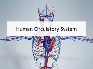
The Human Circulatory System. Introduction. Humans and other vertebrates have a closed circulatory system: This means that circulating blood is pumped through a system of vessels This system consists of the heart (pump), series of blood vessels and the blood that flows through them.

E N D
Introduction • Humans and other vertebrates have a closed circulatory system: • This means that circulating blood is pumped through a system of vessels • This system consists of the heart (pump), series of blood vessels and the blood that flows through them.
The Heart • Located near the center of your chest • Hollow structure • Composed almost entirely of muscle • About the size of your clenched fist • Enclosed in a protective sac called the pericardium
The Heart • In the walls of the heart, two layers of tissue form a sandwich around a thick layer of muscle called the myocardium. • Contractions of the myocardium pump blood through the circulatory system. • The heart contracts about 72 times per minute • Pumps about 70mL of blood with each contraction. • The right and left sides of the heart are separated by a septum, or wall. • The septum prevents the mixing of oxygen rich and oxygen poor blood.
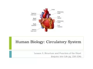
The Heart • On each side of the septum are two chambers. • The upper chamber (receives blood) is the atrium. • The lower chamber (pumps blood out of heart) is the ventricle. • The heart has a total of 4 chambers: • 2 atriums • 2 ventricles
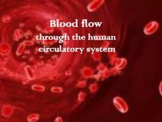
Pathway of Blood • Deoxygenated blood passes from the right atrium into the right ventricle and then goes to the lungs. • From the lungs, blood moves back toward the heart into the left atrium to the left ventricle and then passes into the aorta to go to the rest of the body
Valves • As the heart contracts, blood flows into the ventricles and then out through the ventricles. • Flaps of connective tissue, called valves, are located between the atria and ventricles. • Blood moving keeps the valves open. • When the ventricles contract, the valves close which prevent blood from flowing back into the atria. • There are also valves that stop blood from re-entering the ventricles after the blood has left. • This system of valves keeps blood moving in one direction which increases the pumping efficiency of the heart.
Heart Beat • Heart muscles are composed of individual fibers • Each atrium and ventricle contracts as a unit. • Each contraction begins with a group of cardiac muscle cells in the right atrium known as the senatorial node (SA node) • Because the SA node paces the heart it is known as the pacemaker. • The impulse spreads from the pacemaker to the rest of the atria. • From the atria, a signal is sent to the atrioventricular node and then to a bundle of fibers in the ventricle. • When the ventricle contracts, blood flows out.
Blood Vessels • As blood moves through the circulatory system it moves through 3 types of blood vessels: • Arteries • Capillaries • Veins
Arteries • Large vessels • Carry blood from heart to tissues of body • Carry oxygen rich blood, with the exception of pulmonary arteries. • Thick walls-need to withstand pressure produced when heart pushes blood into them.
Capillaries • Smallest blood vessels • Walls are only one cell thick and very narrow. • Important for bringing nutrients and oxygen to tissues and absorbing CO2 and other waste products.
Veins • Once blood has passed through the capillary systems it must be returned to the heart. • Done by veins • Walls contains connective tissue and smooth muscle. • Largest veins contain one way valves that keep blood flowing toward heart. • Many found near skeletal muscles. When muscles contract, blood is forced through veins.
Blood Pressure • The heart produces pressure • The force of blood on the wall of the arteries is known as blood pressure. • Blood pressure decreases as the heart relaxes, but the rest of the circulatory system is still under pressure.
Blood Pressure • When blood pressure is taken, the cuff is wrapped around the upper portion of the arm and pumped with air until blood flow in the artery is blocked. • As the pressure in the cuff is relaxed, 2 numbers are recorded. • Systolic pressure- the first number taken, is the force felt in the arteries when the ventricles contract. • Diastolic pressure- the second number taken, is the force of the blood on the arteries when the ventricles relax.
Disorders of Circulatory System • Atherosclerosis • Fatty deposits (plaque) in walls of arteries • Deposits can obstruct flow of blood which can raise blood pressure • Increases risk of blood clots • If clot breaks free it can obstruct blood flow to tissues.
Disorders of Circulatory System • Heart Attack • Due to atherosclerosis, coronary arteries may become blocked (blood can’t get to heart muscle) • Heart muscle begins to die due to lack of O2
Disorders of Circulatory System • Stroke • Blood clot may break free and block a vessel leading to the brain. • Brain cells are starved of oxygen and nutrients • Loss of function may occur • Can cause paralysis, loss of ability to speak or death.
Blood • Composed of plasma and blood cells • Types of Cells are: • Red Blood Cells • White Blood Cells • Platelets
Blood • Plasma • Straw colored • 90% water • 10% dissolved gases, salts, nutrients, enzymes, hormones, wastes, and proteins.
Blood • Plasma proteins • 3 Types: Albumins, globulins and fibrinogen. • Albumins and Globulins- transport substances such as fatty acids, hormones and vitamins. • Fibrinogen- Responsible for blood’s ability to clot
Blood • Red Blood Cells • Most numerous type • Transport oxygen • Get color from hemoglobin • Disk shaped • Made in red bone marrow • Circulate for 120 days
Blood • White Blood Cells • Guard against infection, fight parasites, and attack bacteria • Number of WBC’s increases when body is fighting • Lymphocytes produce antibodies which fight pathogens and remember them
Blood • Platelets • Aid the body in clotting • Small fragments • Stick to edges of broken blood cell and secrete clotting factor to help form clot.
Blood Clotting Problems • Hemophelia • Genetic disorder that disrupts clotting • People must be very careful to avoid injury • Can be treated by injecting extracts that contain the missing clotting factor.
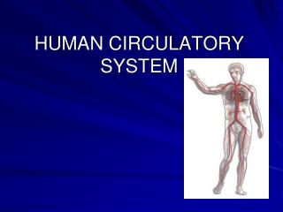
INTRODUCTION • The cardiovascular system is transport system of body • It comprises blood, heart and blood vessels. • The system supplies nutrients to and remove waste products from various tissue of body. • The conveying media is liquid in form of blood which flows in close tubular system.
FUNCTION OF CARDIOVASCULAR SYSTEM • Transport nutrients, hormones • Remove waste products • Gaseous exchange • Immunity • Blood vessels transport blood • Carries oxygen and carbon dioxide • Also carries nutrients and wastes • Heart pumps blood through blood vessels
COMPONENTS OF CARDIOVASCULAR SYSTEM BLOOD HEART BLOOD VESSELS
BLOOD • The Blood: Blood cells & Plasma • Blood cells 1- Erythrocytes - Red Blood Cells 2- Leucocytes 3- Thrombocytes • Plasma is fluid portion
HEART • Heart is a four chambered, hollow muscular organ approximately the size of your fist • Location: • Superior surface of diaphragm • Left of the midline • Anterior to the vertebral column, posterior to the sternum
FUNCTIONS OF THE HEART • Generating blood pressure • Routing blood Heart separates pulmonary and systemic circulations • Ensuring one-way blood flow Heart valves ensure one-way flow • Regulating blood supply Changes in contraction rate and force match blood delivery to changing metabolic needs
BLOOD VESSELS Blood Vessels -A closed network of tubes These includes: Arteries Capillaries Veins
BLOOD VESSELS Arteries(Distributing channel) • Thick walled tubes • Elastic Fibers • Circular Smooth Muscle • Capillaries (microscopic vessels) • One cell thick • Serves the Respiratory System Veins (draining channel) • General structure 1.Tunica intima 2.Tunica media 3.Tunica adventitia
CLASSIFICATION OF BLOOD VESSELS Conducting Vessels Distributing Vessels Resistance Vessels Exchange Vessels Capacitance / Reservoir Vessels
ARTERIES Blood vessels that carry blood away from the heart are called arteries. They are the thickest blood vessels and they carry blood high in oxygen known as oxygenated blood (oxygen rich blood). Accompanied by vein and nerves Lumen is small No valves Repeated branching
CLASSIFICATION OF ARTEIES Elastic- e.g. (Aorta & its Major branches) Muscular -e.g.(Renal, Testicular, Radial, Tibial etc.) Arterioles (<0.1 mm)- Terminal arterioles, Meta-arterioles, Thoroughfare, channel/ preferred
CAPILLARIES (5-8 micron) The smallest blood vessels are capillaries and they connect the arteries and veins. This is where the exchange of nutrients and gases occurs. BODY CONTAINS TWO KINDS OF CAPILLARIES CONTINUOUS-SKIN, LUNG, SMMOTH MUSCLE, CONNECTIVE TISSUES FENESTRATED- PANCREAS,ENDOCRINE GLANDS, SMALL INTESTINE,CHOROID PLEXUS,CILLIARY PROCESS etc.
SINUSOIDS SINUSOIDS- Large irregular vascular space (30-40 micron) eg.Liver, Spleen, Bone marrow, suprarenal, Parathyroid etc.
VEINS Blood vessels that carry blood back to the heart are called veins. They have one-way valves which prevent blood from flowing backwards. They carry blood that is high in carbon dioxide known as deoxygenated blood (oxygen poor blood).
VEINS • Thin Walled • Large irregular lumen • Have valves • Dead space around • Types: Large Medium Small • Veins without valves: • SVC & IVC • Hepatic, Renal • Uterine, Ovarian not Testicular • Facial • Pulmonary • Umbilical • Emissary • Portal Veins <2mm
VEINS • Veins without Muscular tissue: • Dural venous sinuses • Pial Veins • Retinal • Veins of erectile tissue of sex organs • Veins of spongy bones • Factors responsible for venous return: • Muscle contraction • Negative intrathoracic pressure • Pulsation of arteries • Gravity • Valves
ANASTOMOSIS Communication between vessels ARTERIAL: Actual( end to end & convergent)-Palmar, plantar, Circle of Willis, Labial Intestinal arcade, etc. Potential-Coronary, around joints etc.
ANASTOMOSIS ARTERIOVENOUS ANASTOMOSIS: Skin of nose Lips External Ear Mucus membrane of GI & nose Erectile tissue of sex organ Thyroid Tongue
END ARTERIES END ARTERIES: Central artery of retina Arteries of spleen, liver, kidneys, metaphyses of long bones Central branches of cerebral cortex
CIRCULATION • Coronary circulation – the circulation of blood within the heart. • Pulmonary circulation – the flow of blood between the heart and lungs. • Systemic circulation –the flow of blood between the heart and the cells of the body. • Fetal Circulation
SYSTEMIC AND PULMONARY CIRCULATION Pulmonary circulation The flow of blood between the heart and lungs. Systemic circulation The flow of blood between the heart and the cells of the body.
PORTAL CIRCULATION Portal circulation - the flow of blood between tow set of capillaries before draining in systemic veins.