Download
1 / 35
430 likes | 734 Views
Integumentary System. SKIN LAYERS (Epidermis and Dermis) EPIDERMIS (outside to inside) Stratum Corneum Thin and flat cells Dead Continually shed (35 days to new skin) Cytoplasm – full of protein keratin Water repellent Cells are very dry

E N D
SKIN LAYERS (Epidermis and Dermis) • EPIDERMIS (outside to inside) • Stratum Corneum • Thin and flat cells • Dead • Continually shed (35 days to new skin) • Cytoplasm – full of protein keratin • Water repellent • Cells are very dry • If soak in water, cells act as sponge and absorb water • Skin puckers, bulges when in water a long time • Allowed to dry out wrinkles disappear • Barrier to water loss, outside chemicals, and outside bacteria
Stratum Lucidum • Clear layer • Dead - No nuclei • Prevents water loss • Apparent in “thick layers” of skin – soles of feet and palms of hand • Stratum Granulosum • Keratinization begins in this layer • Intense staining granules
Stratum spinosum • Irregular-shaped cells • Desmosomes – joining adjacent cells • Spot welds • Heat, friction, or irritants break bonds and results in blisters • Gives a spiny appearance • Heavy production of keratin (heavy RNA and protein synthesis)
Stratum basale • Single layer of columnar cells • Deepest cells undergo mitosis • Migrate from this layer to surface of skin • Regeneration of cells about 3-4 weeks • Abrasions or constant pressure increases mitotic activity • Leads to a thickened corneum (calluses)
DERMIS – Corium (much thicker than the epidermis) • Thicker than Epidermis • Skin strength in the dermis • Storage area for water
Dermis • Papillary layer • Thin layer that projects into the epidermis – dermal papillae • Responsible for fingerprinting
Dermis • Reticular layer • Collagen fibers give strength to the skin • Commercial processing of animal skin – leather • Elastic fibers give skin its give • Point of attachment for muscle • Results in facial expressions and scalp movement • Hair follicles have arrectorpili muscle attached • Hair stands on end in extreme fright (skin crawl) • Raises the skin around it – “goose bumps” • Doesn’t shed and regenerate • Contains sensory receptors (pain, pressure, touch, and temperature)
SUBCUTANEOUS LAYER OR HYPODERMIS • Not really part of the skin itself • Lies between dermis and underlying tissues • Rich in fat or adipose tissue • Choice for subcutaneous injections and absorption of materials
SKIN COLOR • Determined by 3 factors • Amount of melanin • Carotene – yellow pigment • Hemoglobin at the surface
Melanin – production occurs in Melanocytes • Located in the Stratum basale • Melanin – dark brown pigment • Tyrosine (aa) → Melanin • Requires enzyme tyrosinase • Absence of enzyme – results in albinism • Decreased enzyme – results in graying of hair and white skin of elderly • Controlled by genes • Environmental control – exposure to sun (increases melanin production) • Meant as protection to UV damaging rays (mutations)
Carotene – yellowish color • Hemoglobin – reddish color • Depends on volume of blood in skin capillaries
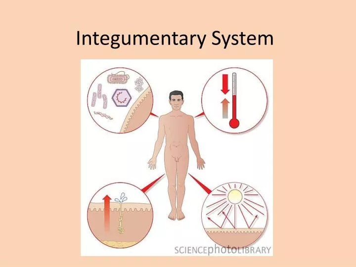
FUNCTIONS OF THE SKIN • Protection – • Microorganisms – mechanical and lysozymes • From water loss – keratin • Chemicals – keratin • Physical damage – Collagen strength • UV radiation - Melanin
Sensation – sensory receptors puts us in contact with our external environment • Pressure, touch, pain, and temperature
Excretion – salts and wastes (ammonia and urea) • Vitamin D production (hormone or endocrine function)– UV light causes a chemical reaction that allows vitamin D formation • Important in calcium absorption for strong bones
Immunity • Macrophages are in the tissue to repel foreign invaders • Langerhans cells work with Helper T cells – trigger immune reaction to disease
Body Temperature Homeostasis (balance) • Heat Loss • Increases the flow of blood to the skin dermis – Vasodilation • Sweating and evaporation allow heat to be removed • Heat Conservation • Decreases the flow of blood to the skin dermis – Vasoconstriction
SKIN APPENDAGES • HAIR • Present all over body EXCEPT – lips, nipples, palms of hands and soles of feet • Parts of the hair • Shaft – hair seen above the surface of the skin • Root part of shaft below the surface • Follicle – tube that surrounds the hair below the surface of the skin • Epidermal layers spread down into the dermis • Contain mitotic layer for hair shaft formation • Melanocytes are present that give color to the hair • Papilla - Supplies blood capillaries for nourishment
Hair color • Amount of melanin deposited in the middle of the hair determines color • Special melanin containing iron results in red hair • Gray and White hair decrease or absence of melanin • Straight or curly hair • Straight hair – round shaft • Curly hair – flat shaft
NAILS • Nail body - visible part of nail (keratinized epithelial cells) • Root of nail – hidden by the cuticle • Lunula - crescent-shaped white area • Stratum germinativum below lunula – site of nail growth • Rich blood vessels below nail give a pink color to nail
SKIN GLANDS (Cutaneous glands) • All are exocrine glands (they release their secretions to the skin surface via ducts) • Sebaceous (Oil) Glands- found all over the skin, except on palms of the hands and soles of the feet. • Sebum- secreted by sebaceous glands • Made up of an oily substance and fragmented cells • A lubricant that keeps the skin soft and moist and prevents the hair from becoming brittle. • Contains chemicals to kill bacteria
Sweat glands- “sudoriferous glands”- more than 2.5 million per person • Eccrine glands- found all over the skin • Produce sweat (water plus some salts, vitamin C, traces of metabolic wastes (ammonia, urea, uric acid), and lactic acid. • The sweat glands are supplied with nerve endings
Apocrine sweat glands- axillary and genital areas • Larger than eccrine glands • Secretions contain fatty acids and proteins as well as all substances present in eccrine secretions. • May be milky or yellow in color. • Odorless, but when bacteria begin to grow on the nutrients in the secretions, it takes on a musky smell. • Apocrine glands begin to function during puberty under the influence of androgen