Download
1 / 21
210 likes | 433 Views
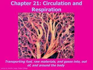
Circulation and Respiration. A.P. Biology. Circulation. Why are circulatory systems necessary in most cases? Single cells, hydra, jellyfish – 2 cell layers thick so can get food and oxygen to cells by direct diffusion – but otherwise it is too slow over long distances.

E N D
Circulation and Respiration A.P. Biology
Circulation • Why are circulatory systems necessary in most cases? • Single cells, hydra, jellyfish – 2 cell layers thick so can get food and oxygen to cells by direct diffusion – but otherwise it is too slow over long distances. • Open circulatory systems – insects, arachnids, most mollusks • Have a heart – usually tubular – pumps to spaces – body movement squeezes the cavities forcing the hemolymph back to the heart • Closed circulatory system – earthworms to humans • Separate blood and interstitial fluid (blood stays in the vessels) • Heart – arteries – arterioles – capillaries (components of blood leave the vessels and enter the interstitial fluid surrounding the cells) - venules - veins
Blood Vessel Structure • Basic blood vessel structure: • Internal lining • Epithelium • Middle of wall • Smooth muscle • Outside • Fibrous connective tissue Capillaries – just one single cell layer thick for diffusion Epithelium connective Smooth muscle
Arteries More smooth muscle Smaller lumen due to pressure A lot of pressure No valves Veins Little smooth muscle Bigger lumen due to less muscle Low pressure Valves Arteries vs. Veins • Veins are under low pressure so it is difficult for blood to get back to the heart from the feet – need help: • Valves prevent backflow • Skeletal muscle helps push blood back • Pressure in chest from breathing
Heart Structure • 2 chambered heart – fish • Heart – gills – body • Lose pressure when go thru gills • 3 chambered heart – amphibians and most reptiles • Have mixing of oxygenated and unoxygenated blood • 4 chambered heart – birds and mammals • Coincides with endotherms
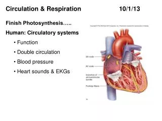
Path of Blood Through the Heart • Rt. Atrium receives oxygen poor blood from the vena cava. • Flows thru to rt. ventrical (thru the rt. AV valve) and pumps to lungs thru pulmonary arteries (thru pulmonary semilunar). • Left atrium receives oxygen rich blood from the lungs thru pulmonary veins. • Blood flows to left ventricle (thru left AV valve) and is pumped out to body through the aorta (thru aortic semilunar valve). • Blood circulates to the heart thru coronary arteries - only when it is relaxed
Electricity Thru the Heart • SA (sinoatrial node – pacemaker) gets the signal from the brain and sends the electrical impulse through the atria to the AV (atrioventricular) node. The atria then contract. • AV node delays the impulse – allows atria to finish contracting • AV node sends the pulse down the AV bundle to the bundle branches to the Purkinje fibers in the walls of the ventricles • Heart contracts from the apex up sending blood up and out thru the aorta and pulmonary artery
Movement of Blood Components In and Out of Blood Vessels • Movement occurs only thru capillaries • Nutrients will move out due to conc. differences (high in bv, low interstitial fluid) • Water moves out at beginning of capillary even though water conc. is higher interstitial fluid because blood pressure overcomes osmotic pressure • At end of capillary, blood pressure has dropped so water reenters capillary • Any extra that doesn’t reenter capillaries, will be picked up by lymph
Respiratory Method Protozoa – cell surface Sponge, cnidarians, flatworms – cell surface – all cell in contact with environment Earthworms, some amphibians – gas exchange across moist body Insects - tracheae Circulatory System No circulatory system No circulatory system Closed circulatory system Open cirulatory system that doesn’t transport Oxygen Respiration – need a large moist surface for oxygen diffusion
Fish – gills Amphibians, reptiles, birds, mammals - all lungs Closed circulatory system, 2 chambered heart Closed, 3 chambered heart Closed, 4 chambered heart Respiratory Systems Continued
Respiratory Surfaces Continued • Gills – outfoldings to increase surface area, no worry about drying out but water is low in oxygen content • Water flows over the gills – some fish have adaptations to move water faster over the gills (ventillation) • Blood flows in opposite direction from the water over the gills – called countercurrent exchange • Trachae – greater concentration of oxygen in air (diffusion is quicker) • Respiratory surface can dehydrate so its folded inside • Internal tubes that branch and go to the cells • Tubes end at moist epithelium where gas exchange happens
Path of Air Thru the Respiratory System • Nasal Cavity → Pharynx → Glottis → Larynx → Trachea → Bronchi → Bronchioles → Alveoli (respiratory surface) • Mucous traps dust and junk and move it up to the pharynx where it can swallowed
Breathing • Diaphragm contracts (pulls down) • Increases the volume of the lungs which decreases the pressure in the lungs • Atmospheric pressure is greater so air flows into the lungs • Intercostal muscles also expand the ribs which increases the chest cavity more and decreases pressure more, helping air to rush in
Breathing • Tidal Volume – normal volume of air inhaled and exhaled due to negative pressure (.5 L) • Vital Capacity – maximum amount of air that the lungs can hold (3.5-4.5 L) • Residual Volume – amount of air left in the lungs after a normal breath out
Breathing Control • Pons, Medulla Oblongata (Brain Stem) • Mostly controlled by CO2 thru pH • A drop in pH in blood in the brain signals the medulla to increase breathing to get rid of CO2 • In extremes, a lack of O2 is picked up by sensors in the aorta and carotid arteries – send information to medulla to increase breathing rate • Stretch centers in lungs send information back to medulla which inhibits further expansion
Loading and Unloading of O2 • 760 mmHg is the pressure exerted by the air • 21% of the air is oxygen so the ppO2 is .21 x 760 = 160 mmHg • The ppO2 of venous blood entering the lungs is ~40 mmHg so O2 diffuses quickly from the air sacs into the blood • In mollusks + arthropods – they have hemocyanin where Cu+ is the binding pigment – blue – oxygen binds to that is more efficiently carried since O2 isn’t very soluble in water • In higher animals, O2 is picked up by hemoglobin that has 4 O2 binding sites with Fe+3 in the sites where the O2 binds
HemoglobinWhy is hemoglobin good at binding O2 and then letting it go where it’s needed? How does tissue most in need get the most O2? • Shape of Hemoglobin is affected by CO2 concentration • Shape of Hemoglobin is affected by O2 concentration • Hemoglobin exhibits cooperativity • Diffusion rates increase with increasing concentration differentials
HemoglobinWhy is hemoglobin good at binding O2 and then letting it go where it’s needed? How does tissue most in need get the most O2? • Hemoglobin changes shape in slightly acidic environments, causing it to dump O2 faster • In high concentrations of O2 like that of the lung – hemoglobin is fully saturated; very low O2 concentrations cause quicker unloading (see dissociation curve) • Cooperativity: Once O2 binds to one of the 4 binding sites, the other sites change shape slightly allowing the other 3 sites to grab onto O2 quicker • When unloading – when 1 leaves, the other 3 leave faster • Once hemoglobin lets go of O2 - Where there is a greater concentration gradient, the faster O2 will diffuse out of the blood.
How CO2 is Carried in the Blood • A little is carried in the plasma (7%) • Some is bound to hemoglobin (23%) • The majority is carried as bicarbonate: (70%) • diffuses into RBC’s and is converted to bicarbonate and H+ • H+ is carried by hemoglobin and the bicarbonate diffuses into the plasma • In the lung, the bicarbonate diffuses back into the RBC ; hemoglobin releases CO2 and H+ which recombines with bicarbonate and forms CO2 again