Download
1 / 24

E N D
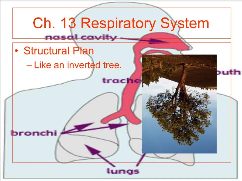
1. Ch. 13 Respiratory System Structural Plan
Like an inverted tree.
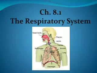
2. Ch. 13 Respiratory System Upper respiratory tract
Nose pharynx and larynx
3. Ch. 13 Respiratory System Lower respiratory tract
Trachea, bronchial tree, lungs
4. Ch. 13 Respiratory System Respiratory mucosa
Specialized membrane that lines tubes of respiratory tree.
More than 125 ml of mucous produced daily
Air purification system by trapping inspired irritants.
Cilia on mucosal cells brat in only one direction,--- upward.
5. Ch. 13 Respiratory System Nose
Structure
Nasal septum separates nose into 2 cavities.
Mucous membrane lines nose
Sinuses drain into nose
Function
Warms and moistens inhaled air
Contains organs of smell.
6. Ch. 13 Respiratory System Pharynx
Structure
Pharynx (throat) is approx. 5 in. long.
Divided into nasopharynx, oropharynx & laryngopharynx.
2 nasal cavities, mouth esophagus, larynx auditory tubes open into the pharynx.
Pharyngeal tonsils and openings of auditory tubes open into nasopharynx, tonsils found into oropharynx
7. Ch. 13 Respiratory System Function
Passageway for food and liquids
Air distribution; passageway for air
8. Ch. 13 Respiratory System Trachea
Structure
Tube approx 4-5 in. long
Has mucous lining
C-shaped cartilage rinds hold trachea open
9. Ch. 13 Respiratory System Function
Passageway for air to move to and from the lungs
Obstruction
Blockage of trachea obstructs air and can cause death in minutes
4000 U.S. deaths annually
Heimlich maneuver used to free trachea of obstruction
10. Ch. 13 Respiratory System Bronchi, bronchioles and alveoli
Trachea breaks into right and left bronchi
Each bronchus branches into smaller bronchioles
Bronchioles end in clusters of microscopic alveolar sacs, made up of alveoli
11. Ch. 13 Respiratory System Function
Bronchi and bronchioles � air distribution; passageway for air to move to and from alveoli.
Alveoli � exchange of gases between air and blood.
12. Ch. 13 Respiratory System Lungs and pleura
Structure
Large enough to fill chest cavity, except for mediastinum that is occupied by the heart and large blood vessels.
Apex - is upper part of each lung under collarbone
Base � broad lower part resting on diaphragm.
Pleura � moist smooth slippery membrane that lines chest cavity and covers surface of each lung. Reduces friction between lungs and chest wall.
13. Ch. 13 Respiratory System Function
Breathing (pulmonary ventilation)
14. Ch. 13 Respiratory System Respiration
Mechanics of breathing
Breathing includes 2 phases, inspiration (air into lungs) and expiration (air out of lungs).
Changes in shape and size of thorax causes changes air pressure within lungs.
Air pressure differences causes air to move in and out of lungs.
15. Ch. 13 Respiratory System Inspiration
Active process � air moves into lungs
Inspiratory muscles include diaphragm and external intercostals.
Diaphragm muscle flattens during inspiration, increases top to bottom length of thorax.
External intercostals contraction elevates the size of the thorax from front to back and from side to side
The increase of the size of the chest cavity reduces pressure within it, and air enters the lungs.
16. Ch. 13 Respiratory System Expiration
Quiet expiration is a passive process.
During expiration, thorax returns to original shape and size.
Elastic recoil of lung tissue aids in expiration
Expiratory muscles used in forceful expiration are internal intercostals and abdominal muscles
Internal intercostals, compresses rib cage and decreases size of thorax from front to back
Contraction of abdominals elevated diaphragm decreasing size from top to bottom
Reduction in size of thoracic cavity increases pressure and air leaves lungs.
17. Ch. 13 Respiratory System Exchange of gases in lungs (figure 13-12)
Carbaminohemoglobin breaks down into CO2 and hemoglobin.
CO2 move out of lung capillary blood into alveolar air and out of the body in expired air.
Oxygen moves from alveoli into lung capillaries
Hemoglobin combines with oxygen to form oxyhemoglobin.
18. Ch. 13 Respiratory System Exchange of gases in tissues
Oxyhemoglobin breaks down into O2 and hemoglobin
O2 moves out of tissue capillary blood into tissue cells.
CO2 moves from tissue cells into tissue capillary blood
Hemoglobin combines with CO2 forming Carbaminohemoglobin
19. Ch. 13 Respiratory System Volumes of air exchanged in breathing
Can be measured with spirometer
Tidal volume � amount breathed in or out with each breath
Vital capacity � greatest amount that can be breathed in out on one breath
Expiratory reserve volume � amount of air forcibly exhaled after expiring tidal volume
Inspiratory reserve volume � amount of air forcibly inhaled after normal inspiration
Residual volume � air that remains in lungs after the most forceful expiration
Rate � 12 � 18 breaths per minute, increased during exercise.
20. Ch. 13 Respiratory System Regulation of respiration
Regulation of respiration permits body to adjust to varying demands for O2 supply and CO2 removal
Most important regulatory centers on medulla are called respiratory control centers.
Under resting conditions, nervous activity in the respiratory control centers produces a normal rate and depth of respirations (12 to 18 per minute)
21. Ch. 13 Respiratory System Control of respiration
control home
22. Ch. 13 Respiratory System Respiratory control centers in the medulla are influenced by �inputs� from receptors located in other body areas:
Cerebral cortex � voluntary, but limited control of respiratory activity
Chemoreceptors respond to changes in CO2 and O2 and blood acid levels � located in carotid and aortic bodies
Pulmonary stretch receptors � respond to the stretch of lungs, thus protecting respiratory organs from over inflation
23. Ch. 13 Respiratory System Carotid and arotic bodies
24. Ch. 13 Respiratory System Types of breathing
Eupnea � normal breathing
Hyperventilation � rapid and deep respirations
Hypoventilation � slow and shallow respiration
Dyspnea � labored or difficult respirations
Apnea � stopped breathing
Respiratory arrest � failure to resume breathing