Download
1 / 1
30 likes | 172 Views
Recollection and the Hippocampus. Recollection-Based Memory in Frontotemporal Dementia. Frontal Battery and Recollective Memory. Recollection and the Prefrontal Cortex. Much evidence links the hippocampus with recollection-based memory. For example:. Study.

E N D
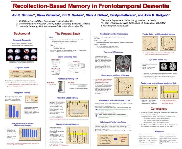
Recollection and the Hippocampus Recollection-Based Memory in Frontotemporal Dementia Frontal Battery and Recollective Memory Recollection and the Prefrontal Cortex Much evidence links the hippocampus with recollection-based memory. For example: Study Currently no volumetric protocol for the prefrontal cortex, so two methods of testing this: Semantic Dementia Control Mean Source Discrimination 100 Associative Memory Test Vargha-Khadem et al. (1997): • Schacter et al. (1984), Glisky et al. (1995): 90 Source memory correlates highly with scores on frontal lobe tests in healthy adults. 80 Selective bilateral hippocampal damage impairs recall but spares recognition memory Percent Correct 70 • Temporal variant of Frontotemporal dementia • Progressive atrophy of one or both temporal lobes Key: Compare recollection-based memory with performance on frontal lobe tests in patients with semantic dementia. Jon S. Simons1*, Mieke Verfaellie2, Kim S. Graham1, Clare J. Galton3, Karalyn Patterson1, and John R. Hodges1,3 Aggleton & Brown (1999): 60 Control group confidence interval Patient confidence interval (no overlap indicates impairment) r=.72, p<.05 r=.69, p=.056 50 • ‘Recollection’ supported by hippocampal system • ‘Familiarity’ supported by perirhinal cortex system 40 Composite Frontal Score Composite Frontal Score • Schacter et al. (1984), Shimamura et al. (1990): 30 Source memory impairment prominent feature of frontal lobe damage. GCB KH DM MB DC JH DE FM Patients with Semantic Dementia Patients (increasing severity ) Patients (increasing severity ) Examine source memory ability of patients with the frontal variant of frontotemporal dementia. * Now at the Department of Psychology, Harvard University, 1. MRC Cognition and Brain Sciences Unit, Cambridge, UK 2. Memory Disorders Research Center, Boston University School of Medicine 3. University Neurology Unit, Addenbrooke’s Hospital, Cambridge, UK • Composite frontal score correlates significantly with both source discrimination and associative memory. Rm 860, William James Hall, 33 Kirkland St, Cambridge, MA 02138 E-mail: jss@wjh.harvard.edu Volumetric MRI Analysis Source Monitoring Task 1) Battery of Frontal Lobe Tasks 2) Frontal Variant FTD • Tests chosen to reflect different aspects of frontal lobe function, such as temporal sequencing, planning, holding and manipulating within working memory. • Z-scores for each individual patient calculated for each frontal test, relative to mean performance of healthy controls. • Composite frontal score then computed for each patient by taking the average of the frontal test z-scores. Volumetric analysis of the hippocampus (and other temporal lobe regions) conducted by manually tracing on 1.5mm contiguous coronal MRI slices using Analyze software on a Sun Sparcstation 20. Areas totalled to produce a volume for each region, corrected for total brain volume by dividing by whole brain cross-sectional area. Cognitive Profile Regions of significant gray matter density reduction relative to age-matched controls, as measured by voxel-based morphometry (Mummery et al., 2000). Impaired Preserved Study Phase 1 Study Phase 2 Semantic Memory (all modalities of input and output) Phonology, syntax Problem solving Visuospatial Perception • Frontal variant of Frontotemporal dementia • Progressive atrophy primarily affecting frontal lobes 10 min delay Test Phase • Wisconsin Card Sorting Test • Tower of London • WMS-R Digit Span • WMS-R Spatial Span • Computerized Spatial Span • Computerized N-Back 30 different drawings 30 line drawings Composite Frontal Score 120 drawings: 30 from Set 1, 30 Set 2, 60 New • Earliest signs are changes in behavior and personality, followed by cognitive impairment (attention, executive function, etc.) • Recognition memory deficit rarely observed, unless atrophy has spread to involve posterior cortical regions “Did you see the picture in Set 1, Set 2, or not at all?” Recent experiments suggest better retrieval of recent autobiographical memories compared to those from the distant past (e.g., Graham & Hodges, 1997). Associative Memory Test • This implies that learning of new material may be relatively preserved in semantic dementia Performance on the Source Monitoring Task Study Phase 32 door and sofa pairs Item Detection Recognition Memory We have investigated episodic learning in semantic dementia using objects, faces, and line drawings as stimuli (Graham et al., 2000; Simons et al., 2001). Test Phase • Patients with frontal variant FTD had little difficulty with familiarity-based item memory. • But all patients tested were severely impaired at recollection-based source memory. 48 door and sofa pairs: 16 old, paired together at study 16 old, re-paired since study 16 new, not seen at study “Were these items paired together at study?” Item Detection Typically, patients with semantic dementia (SD) show intact recognition memory for these kinds of pictures. Patients with early Alzheimer’s disease (AD), however, exhibit marked impairment. Mean No. Correct * Conclusions Source Discrimination • Item memory normal in most patients with semantic dementia; the most severe patients may be impaired. • Source discrimination normal in some patients, impaired in others. • Source memory not related to bilateral hippocampal volume • Strong association with performance on frontal lobe tasks • Patients with frontal variant FTD very impaired on source • In seeking to understand the neural substrates of long-term memory, we should not forget the important role played by the prefrontal cortex. Analysis of Individual Patients with Semantic Dementia • Most patients with semantic dementia have highly accurate recognition memory. • But at advanced stages of the disease, evidence of emerging deficit? References Source Discrimination Aggleton, J.P. & Brown, M.W. (1999). Behavioral and Brain Sciences, 22, 425-489. Glisky, E.L., Polster, M.R., & Routhieaux, B.C. (1995). Neuropsychology, 9, 229-235. Graham, K.S. & Hodges, J.R. (1997). Neuropsychology, 11, 77-89. Graham, K.S., Simons, J.S., et al. (2000). Neuropsychologia, 38, 313-324. Mandler, G. (1980). Psychological Review, 87, 252-271. Mummery, C.J., Patterson, K., et al. (2000). Annals of Neurology, 47, 36-45. Schacter, D.L., Harbluk, J.L., & McLachlan, D.R. (1984). Journal of Verbal Learning and Verbal Behavior, 23, 593-611. Shimamura, A.P., Janowsky, J.S., & Squire, L.R. (1990). Neuropsychologia, 28, 803-813. Simons, J.S., Graham, K.S., et al. (2001). Neuropsychology, 15, 101-114. Simons, J.S., Graham, K.S., & Hodges, J.R. (submitted). Journal of Memory and Language. Vargha-Khadem, F., Gadian, D.G., et al. (1997). Science, 277, 376-380. * * * What could be causing this recognition memory impairment at late stages of semantic dementia? Background The Present Study Aims: • Investigate relative contributions of recollection and familiarity (Mandler, 1980). • Recollection: ‘Remembering’ item with associated context • Familiarity: ‘Knowing’ that item has been experienced Using: • Source monitoring task • Associative memory test • Relate memory performance to measures of atrophic disruption in associated neural regions (e.g., medial temporal lobe, prefrontal cortex) Hippocampus and Source Memory • No significant positive correlation between bilateral hippocampal volume and source memory. • This is despite significant correlations for other regions (e.g., between inferior temporal lobe and semantic memory). • Suggests that, in semantic dementia at least, hippocampal atrophy is not the explanation for the pattern of recollection-based memory performance. Source Discrimination r=-.4, n.s. HC Volume Z-Score Familiarity-Based Memory • Just like before, as a group, the patients were not impaired relative to controls at familiarity-based item memory, F1,20 = 2.3, n.s. • Also similar to before, three patients at advanced stages of semantic dementia did show a significant deficit. • Previous studies indicate that this pattern is not due to: • Degraded semantic knowledge • Disrupted perceptual processes • But can be attributed to atrophy * * * affecting medial temporal lobe regions such as the perirhinal cortex (Simons et al., submitted). Recollection-Based Memory • Recollection-based source memory of seven patients not significantly impaired relative to controls (although two, WM and IF, are borderline). • Three patients (JP, DS, and JW) significantly impaired. No obvious link between this deficit and disease severity. • Again, pattern not related to degraded semantic knowledge. • Possible explanations? Atrophy progressing to affect the function of: • Hippocampus? • Prefrontal Cortex?