Download
1 / 26
260 likes | 266 Views
Learn about the structure of cells and the process of DNA extraction from blood cells for various purposes such as forensics, evolution, and medical research.

E N D
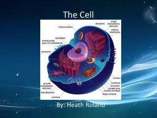
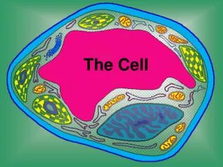
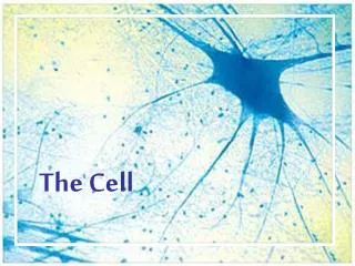
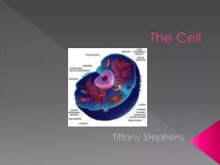
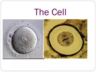
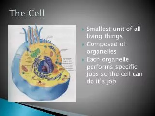
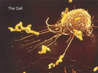
The cell • The basic unit of any living organism. • It contains a complete copy of the organism's genome. • Cells are of many different types (e.g. blood, skin, nerve cells, etc.), but all can be traced back to one special cell, the fertilized egg.
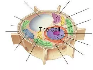
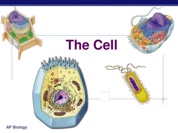
Eukaryotes vs. prokaryotes • Prokaryotic cells: lack a distinct, membrane-bound nucleus. E.g. ?? • Eukaryoticcells: distinct, membrane-bound nucleus. Larger and more complex in structure than prokaryotic cells. E.g. ???
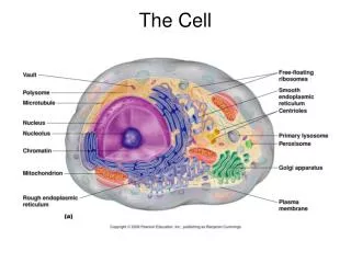
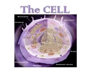
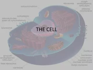
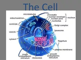
Genetic Material Containing Organelles • Nucleus: The cell nucleus is a membrane bound structure that contains the cell's hereditary information and controls the cell's growth and reproduction. It is the command center of a eukaryotic cell and is commonly the most prominent organelle in a cell. • Mitochondria: Mitochondria are structures within cells that convert the energy from food into a form that cells can use. Although most DNA is packaged in chromosomes within the nucleus, mitochondria also have a small amount of their own DNA. This genetic material is known as mitochondrial DNA or mtDNA. • Chloroplast: It contains its own DNA, which is called chloroplast DNA, abbreviated as cpDNA and also known as plastome. Present in Plants.
DNA Structure • DNA consists of two molecules that are arranged into a ladder-like structure called a Double Helix. • A molecule of DNA is made up of millions of tiny subunits called Nucleotides. • Each nucleotide consists of: • Phosphate group • Pentose sugar • Nitrogenous base
DNA is extracted from human Blood cells for a variety of reasons • Forensics: • Evolution: • Medical: • Bio-engineering or cloning • General research purposes
Before DNA Extraction • Take blood (5ml-10ml) via sterile syringe • Pore blood in to anticoagulant tube or a falcon tube containing 200 micro liter 1M EDTA • Store at -80 for 4,5 hours or -20 for 1 day to provide cold stress to RBC,s • Thaw blood properly before extraction and mix gently
Reagents required • Lysis buffer (10mM Tris HCL, 2mMEDTA, pH 8.0) • TNE buffer (Tris HCL 10 mM, EDTA 2mM, NaCl 400mM) • 10% SDS • Proteinase-K solution 20mg/ml • 6M NaCl • Phenol-Choloroform-Isoamylalcohol(PCI) (25:24:1) • Chilled Isopropanol • 75% Ethanol • Low TE buffer (10mM Tris, 0.2mM EDTA)
Major Steps • Cell lysis and purification of leukocytes • Proteins and Lipid degradation • DNA purification
DNA Extraction (1st day) • Before starting the DNA extraction liquefy or thaw the samples of blood and take 250ul (micro liter)Blood from stock. • Add up to 1 ml of lysis buffer in 1 ml blood containing Eppendorf. • Centrifuge at 6000 rpm for 10 minutes ( All centrifugation in successive phases should be done at 8C)
Remove 250 ulsupernatant. Breakdown the pellet made at lowermost by taping it gradually. Add lysis buffer up to 1ml. • Centrifuge at 6000 rpm for 10 minutes ( All centrifugation in successive phases should be done at 8C) • Remove 500 ulsupernatant. Breakdown the pellet made at lowermost by taping it gradually. Add lysis buffer up to 1 ml. • Centrifuge at 6000 rpm for 10 minutes ( All centrifugation in successive phases should be done at 8C)
Remove 750 ulsupernatant. Breakdown the pellet made at lowermost by taping it gradually. Add lysis buffer up to 1 ml. • Centrifuge at 6000 rpm for 10 minutes ( All centrifugation in successive phases should be done at 8C) • Remove 900 ulsupernatant. Breakdown the pellet made at lowermost by taping it gradually. Add lysis buffer up to 1 ml. • Centrifuge at 6000 rpm for 10 minutes ( All centrifugation in successive phases should be done at 8C)
Remove all supernatant. Breakdown the pellet made at lowermost by taping it gradually. Add lysis buffer up to 1 ml. • Centrifuge at 6000 rpm for 10 minutes ( All centrifugation in successive phases should be done at 8C) • Remove the supernatant leaving pellet and re-suspend pellet in 150 ulTNE buffer for 250 micro liter initial blood volume. Add 20 ul 10% SDS and 2ul Proteinase K. • Samples were incubated overnight in 37dnnn C shakers
Day 2nd • Whole digestion of the pellet is crisscross after one night incubation. If the pellet is not fully digested then add additional Proteinase K according to the quantity of undigested pellet. Once more incubated at 37C for 2-3 hours or till the pellet is fully digested.
Inorganic DNA Extraction Method:1st day protocol is same in both methods: • We preferred inorganic DNA Extraction protocol for fresh samples. • The tubes should kept on snow and added 150 ulsaturated NaCl (6 M) for 250 ul initial blood volume. The falcon tubes were shaken energetically and place on ice for a second time for 10-15 minutes. • Centrifuged at 6000 rpm for 10 minutes to settle down the pellet (salts and proteins). • Supernatant poured in a labeled Eppendorf. • Centrifuged at 6000 rpm for 10 minutes
Organic DNA Extraction Method by Phenol, Chloroform, Isoamylalcohol (PCI): • For old samples we applied Organic DNA Extraction method (PCI). 1st day protocol is same in both methods: • Phenol : Chloroform : Isoamylalcohol • 25 : 24 : 1 • Add 150 ulof PCI for 250 ulinitial blood volume. The falcon tubes were shaken energetically and place on room temperature for 15-20 minutes. • centrifuged at 6000 rpm for 10 minutes to settle down the pellet (salts and proteins). • The supernatant poured in a labeled Eppendorf. • Centrifuged at 6000 rpm for 10 minutes
The same amounts of Isopropanol were added and overturned the tubes moderately until DNA was obvious. • The tubes were leaved for 5 minutes on ice. • Again Centrifuged the samples at 6000 rpm for 10 minutes • The supernatant were thrown away carefully • The DNA pellet was wash away with 150ul of 70 % ethanol for 250 ul initial blood volume.
Centrifuged again at 6000 rpm for 10 minutes • 70 percent ethanol was removed wisely leaving the pellet. • The DNA pellet was air dried out in 37C air dryer. • Added 40 ul low T.E (Tris HCL 10mM, EDTA 0.2mM)/ or in Injection Water • The tubes were placed in 37C shaking incubator overnight to melt the DNA. Covered bands of Para film round the tip of the tubes.
3rd day • Keep the tubes in a shaky water bath 70C for an hour to deactivate nucleases • The tubes were left at room temperature to be chilled • Samples were Spin briefly • 2ml autoclaved tubes were labeled by side and cap. Tubes were labeled including pedigree number individuals ID. • DNA samples were Aliquoted in duplication and stored at -20C conferring to pedigree number in marked and numbered Cryoboxes. • The samples were Stored at -80 C for long term storage