Download
1 / 1
10 likes | 162 Views
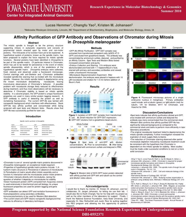
Research Experience in Molecular Biotechnology & Genomics Summer 2010. Center for Integrated Animal Genomics. Lucas Hemmer 1 , Changfu Yao 2 , Kristen M. Johansen 2 1 Nebraska Wesleyan University, Lincoln, NE 2 Department of Biochemistry, Biophysics, and Molecular Biology, Ames, IA.

E N D
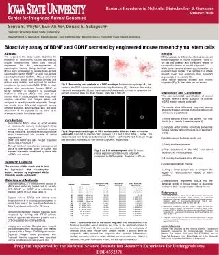
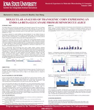
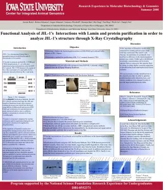
Research Experience in Molecular Biotechnology & Genomics • Summer 2010 Center for Integrated Animal Genomics Lucas Hemmer1, Changfu Yao2, Kristen M. Johansen2 1Nebraska Wesleyan University, Lincoln, NE 2Department of Biochemistry, Biophysics, and Molecular Biology, Ames, IA Affinity Purification of GFP Antibody and Observations of Chromator during Mitosis in Drosophila melanogaster Abstract The mitotic spindle is thought to be the primary structure supporting mitosis in eukaryotic organisms and consists of polymerized tubulin enacted upon by motor and non-motor proteins. The intricacies of its function have yet to be explained. A hypothetical system of proteins, known as the spindle matrix, has been proposed to explain the force required for mitotic spindle functions. Several proteins have been identified in Drosophila to be part of this spindle matrix. Of particular interest is Chromator, which has yet to be tested for persistence in the event of mitotic spindle disassembly, which is one of the predictions of the hypothetical spindle matrix. Drosophila embryos were collected, fixed, stained, and observed under a fluorescent microscope. Control stainings with anti-Skeletor and –Chromator antibodies revealed spindle-like staining that co-incided with the microtubule spindle. However, the mitotic spindle failed to depolymerize using the known microtubule destabilizing nocodazole, leaving inconclusive observations of Chromator’s behavior. A possible problem with nocodazole storage likely reduced its effectiveness during treatment, and thus more observations will be necessary to determine if Chromator stability is based on mitotic spindle stability. In a second project, the GFP protein’s unique fluorescent properties are used in many similar protein tagging research. A GFP antibody is useful in detecting the protein as well as revitalizing fluorescence. The current GFP-Ab was tainted with nonspecific background which interferes with effectiveness. Stock GFP-Ab was affinity purified and concentrated while being analyzed with spot tests and Western blots. Results show that GFP-Ab was successfully purified for later use. • Methods • GFP-Ab Affinity Purification. GFP-GST complex was extracted from transformed competent cells (pGEX-4T-2-GFP) and eluted from glutathione beads. The antibody was concentrated by ammonium sulfate, dialyzed, and purified by an Affinity Column. Spot Tests and Western Blots tested increased concentration and purity. • Embryo Collection and Staining. 3 hr embryos were dechorionated and fixed with 1:1 heptane/Bouin’s fluid, stained with primary and secondary antibodies and viewed under a fluorescent microscope. • Microtubule Depolymerization Experment. After dechorionation, the embryos were placed in heptane with 10 μM nocodazole and shaken for 1.5 min before addition of Bouin’s fluid. A Tubulin Skeletor DNA Composite B Tubulin Chromator DNA Composite CONTROL C Tubulin Skeletor DNA Composite NOCODAZOLE TREATMENT D Chromator Skeletor DNA Composite Results Marker 3rd Elute 2nd Elute 2 hr 1st Elute 3 hr Marker 4 hr 0 hr Figure 4: Fluorescent microscopy pictures of a single embryonic nucleus in metaphase. Primary antibodies used include: anti-α-tubulin (green) or IgM-tubulin (red) for tubulin, 1A1 for Skeletor, 6H11 for Chromator, and Hoechst for DNA. GFP-GST B A • Discussion/Conclusions • Spot tests indicate that affinity purification diluted anti-GFP, once treated with ammonium sulfate and dialyzed the antibody was more concentrated than the commercial stock • Western Blot confirms that affinity-purified GFP-Ab has signficantly reduced background as compared to the commercial stock and looks to perform more efficiently in later laboratory procedures • The original nocodazole treatment failed to depolymerize the mitotic spindle completely. Further investigation revealed the expiration of stock supply likely to blame. • The reduction of Chromator staining observed after partial microtubule depletion induced by nocodazole treatment could be consistant with the hypothesis that Chromator is dependent on the mitotic spindle for stability. More studies must be made under successful conditions to rest on a more solid conclusion Figure 1: Isolation of GFP-GST complex from transformed E. coli. A: Small induction for GFP-GST expression. B: GFP-GST elutions from a glutathione bead column. • Introduction • Chromator is one of several spindle matrix proteins discovered in Drosophila melanogaster, an exceptional model organism • Properties of hypothetical spindle matrix proteins include: 1) A fusiform matrix structure 2) Persistence in absence of microtubules 3) Perturbation of matrix would affect mitotic assembly and or function 4) Interaction with the microtubules and/or motor proteins • Chromator interacts directly with microtubules, raising the question of whether it depends on microtubules for stability • Green fluorescent protein (GFP) extracted from Auquoreavictoriais used extensively in biochemical and cell biology; its unique fluorescent properties are used for protein tagging and gene expression • GFP antibodies can detect GFP and revitalize fluorescence in fixed preparations using secondary antibodies, immunocytochemistry or be used for Western Blot detection • The current stock anti-GFP retains nonspecific background that reduces its efficiency in laboratory techniques Affinity Purified Control 1:1 1:5 1:50 1:100 1:1 1:5 1:50 1:100 A 1:100 1:500 1:1000 1:3000 1:100 1:500 1:1000 1:3000 B Figure 2: Spot tests of anti-GFP with dilution concentrations. A: Prior to concentration. B: After being concentrated with ammonium sulfate and dialyzed in PBS. 1:1000 1:1000 1:100 1:200 1:500 1:50 1:10 • References • Ding Y, Yao C, Lince-Faria M, Rath U, Cai W, Maiato H, Girton J, Johansen KM, Johansen J. 2009. Chromator is required for proper microtubule spindle formations and mitosis in Drosophila. Developmental Biology 334: 253-263. • Johansen J, Johansen KM. 2009. The spindle matrix through the cell cycle in Drosophila. Fly 3:3, 1-8. • Johansen KM, Johansen J. 2007. Cell and molecular biology of the spindle matrix. • International Review of Cytology 263: 157-207 • Johansen KM, Johansen J. 2003. Studying nuclear organization in embryos using antibody tools. Methods in Molecular Biology 247: 215-234. • Qi H, Rath U, Wang D, Xu Y, Ding Y, Zhang W, Blacketer MJ, Paddy MR, Girton J, Johansen J, Johansen KM. 2004. Megator, an essential coiled-coil protein that localizes to the putative spindle matrix during mitosis in Drosophila. Molecular Biology of the Cell 15: 4854-4865. • Rath U, Wang D, Ding Y, Xu Y, Qi H, Blacketer MJ, Girton J, Johansen J, Johansen KM. 2004. Chromator, a novel and essential chromodomain protein interacts directly with the putative spindle matrix protein Skeletor. Journal of Cellular Biochemistry 93: 1033-1047. • Tsien RY. 1998. The Green Fluorescent Protein. Annual Review of Biochemistry 67:509-544. Anti-GFP Commercial anti-GFP Anti-tubulin Figure 3: Western blot of NCD-GFP fusion protein detected with affinity purfied anti-GFP with anti-tubulin as the control and dilution ratios above. Acknowledgements I would like to thank my mentor, Dr. Kristen M. Johansen, and her collaborator, Dr. Jørgen Johansen, for letting me use their lab. I would also like to thank Changfu Yao for his help with my project as well as the other members of the laboratory for support. Finally, I thank the National Science Foundation, Iowa State University, and especially Dr. Max Rothschild and Justin Rice for putting together this REU program and allowing me this opportunity to perform research. Program supported by the National Science Foundation Research Experience for Undergraduates DBI-0552371