Download
1 / 69
1.49k likes | 5.08k Views
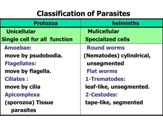
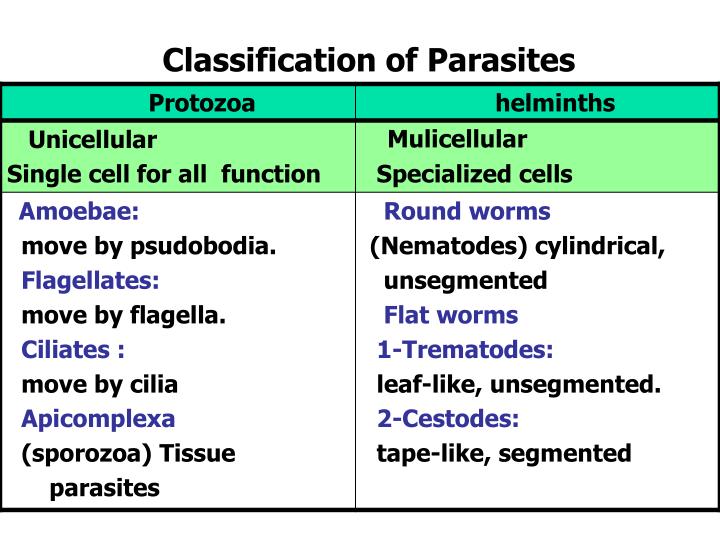
Classification of Parasites. Nematodes General features : Elongated worm, cylindrical, unsegmented and tapering at both ends. Variable in size, measure <1 cm to about 100cm . Sex separate and male is smaller than female. Location of Nematodes: Intestinal nematodes Tissue nematodes .

E N D
Nematodes General features: Elongated worm, cylindrical, unsegmented and tapering at both ends. Variable in size, measure <1 cm to about 100cm. Sex separate and male is smaller than female
Location of Nematodes: Intestinal nematodes Tissue nematodes
Common intestinal nematode infections: Enterobius (Oxyuris) vermicularis(Pinworm,seatworm,threadworm) Trichuris trichiura(whipworm) Ascaris lumbricoides(roundworm) Ancylostoma duodenale & Necator americanus(hookworms) Strongyloides stercoralis :
(Pin worm, seat worm, thread worm( Found all over the world. adult in lumen of cecum and appendix from which adult female migrate to rectum. It can be seen by naked eye as white thread ± 1cm. Male is smaller than female ± 0.5cm, with coiled end. Enterobius vermicularis (Oxyuris)
Enterobius vermicularis (Oxyuris) LIFE CYCLE
Pathology Majority of infectiona are asymptomatic. Main clinical presentation pruritus ani perianal excoriation Ectopic enterobiasis occurs in female when invade valva and vagina result in valvovagintis Usually accompanied by insomnia, anorexia, loss of weight and concentration (Side effect) Enterobius vermicularis (Oxyuris)
Treatment ِِAlbandazole , Mebendazole for whole family Enterobius vermicularis (Oxyuris)
Ascaris lumbricoides (roundworm) Ascaris adult
The commonest human helminthes infection. Found in jejunumand upper part of ileum. Female ± 20 cm longer than male ± 10 cm Feed on semi digested food. Ascaris lumbricoides (roundworm)
Ascaris lumbricoides (roundworm) LIFE CYCLE
Ascaris lumbricoides (roundworm) Ascaris egg (embryonated)
Ascaris eggs Ascaris larva emerging from egg Ascaris egg (embryonated)
Pathology: 1-Adult worm: Light infection : asymptomatic. Heavy infection : intestinal obstruction Migrating adult : to bile duct -jaundice 2-Larvae: Loeffler`s syndrome Pneumonia, cough with bloody sputum Eosinophilia, urticaria Ascaris lumbricoides (roundworm)
Ascaris lumbricoides (roundworm) Loeffler`s syndrome:Larvae in lung pnumonia,cough ,bloody sputum
Ascaris lumbricoides (roundworm) Ascaris larva in lung
Diagnosis: -eggs in stool. -larvae in sputum. -adult may pass with stool. Ascaris lumbricoides (roundworm) Treatment: Albendazole , Mebendazole
Trichuris trichiura (Whipworm) LIFE CYCLE
World wide ,common in poor sanitation. It coexists with Ascaris because of similar requirement. Adult live in large intestine especially caecum and appendix–inheavy infection the whole length of large intestine affected. Male and female worm have narrowanterior portion penetrate the intestinal mucosa Trichuris trichiura (Whipworm)
Pathology light infection : asymptomatic heavy infection :abdominal pain ,bloody diarrhea. Rectal prolapse in children is a common complication. -Diagnosis:egg in stool characterized by its barrel shape with mucoid plugs at each pole . Treatment :Albendazole. Trichuris trichiura (Whipworm)
Trichuris trichiura (Whipworm) Embryonated egg Unembryonated egg Infective stageDiagnostic stage
-Diagnosis:egg in stool characterized by its barrel shape with mucoid plugs at each pole . Treatment :Albendazole. Trichuris trichiura (Whipworm)
Hook worms Ancylostoma dudenale &Necator americanus
1- Buccal cavity with intestinal mucosa 2- B.cavity with teeth &cutting plates anemia
Hook worms Ancylostoma dudenale &Necator americanus LIFE CYCLE
A common cause of anemia. Found in small intestine mainly jejunum. Its buccal capsule (mouth) lined with hard hooks, triangular cutting plates and anticoagulant glands. Hook worms Ancylostoma dudenale &Necator americanus
Hook worms Ancylostoma dudenale &Necator americanus pathology& clinical picture: - larvae: i-At the site of entry of larvae (ground itch). ii- Migration phase: cough with bloody sputum pneumonia, eosinophilia,urticaria. - Adult worm: •low worm burden: no symptoms. •Moderate to heavy burden: epigastric pain, vomiting ,simulating duodenal ulcer, hemorrhagic enteritis.
• Protein loss: hypoproteinaemia edema. •Anemia: due to withdrawal of blood by parasites and hemorrhage from punctured sites lead to sever anemia = microcytic hypochromic . Hook worms Ancylostoma dudenale &Necator americanus
Diagnosis: -Eggs in stools.; -occult blood (+) Hook worms Ancylostoma dudenale &Necator americanus Treatment: Albendazol, Mebendazole
Strongyloides stercoralis Widely distributed in tropical region worldwide . fetal opportunistic in immuno-compromised host. It is smallest pathogenic nematodes ± 2.5mm. adult live in mucous membrane of duodenum jejunum
Strongyloides stercoralis LIFE CYCLE
Pathology and clinical picture: 1-Cutaneouslittle reaction on penetration. sever dermatitis at perianal region in case of external autoinfection. 2- Migration :same as hook worms . 3- Intestinal: inflammation of upper intestinal mucosa, diarrhea, upper abdominal pain clicky in nature. Disseminated strongyloidiasis : in patient with immunodeficiency ,uncontrolled diarrhea –granulomatus changes –necrosis--perforation--peritonitis--death. Strongyloides stercoralis
Diagnosis: rhabditiform larvae diagnostic stage in: -Stool examination -Duodenal aspirate Treatment:Albandazole, Mebendazole Strongyloides stercoralis
TISSUE NEMATODES TISSUE NEMATODES
COMMON TISSUE NEMATODE INFECTIONS • Trichinella spiralis adults in small intestine larvae in tissues (mainly in muscles). • Toxocara canis (dog roundworm) larvae in organs (liver brain eyes), causing visceral larva migrans • Dracunculus medinensis (guinea worm) adult female in subcutaneous tissues • Filarial worms
Most prevalent in areas where domestic pigs are allowed to roam freely.
Trichinosis Pathology: Adults cause mild gastroenteritis. Larvae cause fever, myositis and multi-system involvement which may lead to death. Diagnosis: serology, muscle biopsy. Treatment: Albendazole or Mebendazole + corticosteroids
Visceral larva migrans:Caused by Toxocara canis • Mainly affects children who eat soil contaminated with emberyonated (infective) eggs of Toxocara canis. • Larvae do not develop in humans but migrate continuously in viscera and encapsulate, causing tissue damage.