Download
1 / 36
410 likes | 839 Views
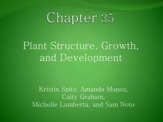
Chapter 35. Plant Structure, Growth, and Development. Figure 35.1 Fanwort (Cabomba caroliniana). Reproductive shoot (flower). Terminal bud. Node. Internode. Terminal bud. Shoot system. Vegetative shoot. Blade Petiole. Leaf. Axillary bud. Stem. Taproot. Root system.

E N D
Chapter 35 Plant Structure, Growth, and Development
Reproductive shoot (flower) Terminal bud Node Internode Terminal bud Shoot system Vegetative shoot Blade Petiole Leaf Axillary bud Stem Taproot Root system Lateral roots Figure 35.2 An overview of a flowering plant
(c) “Strangling” aerialroots (a) Prop roots (b) Storage roots (d) Buttress roots (e) Pneumatophores Figure 35.4 Modified roots
(a) Stolons. Shown here on a strawberry plant, stolons are horizontal stems that grow along the surface. These “runners” enable a plant to reproduce asexually, as plantlets form at nodes along each runner. Storage leaves (d) Rhizomes. The edible base of this ginger plant is an example of a rhizome, a horizontal stem that grows just below the surface or emerges and grows along the surface. Stem Node Root (b) Bulbs. Bulbs are vertical, underground shoots consisting mostly of the enlarged bases of leaves that store food. You can see the many layers of modified leaves attached to the short stem by slicing an onion bulb lengthwise. Rhizome (c) Tubers. Tubers, such as these red potatoes, are enlarged ends of rhizomes specialized for storing food. The “eyes” arranged in a spiral pattern around a potato are clusters of axillary buds that mark the nodes. Root Figure 35.5 Modified stems
(a) Simple leaf. A simple leafis a single, undivided blade.Some simple leaves are deeply lobed, as in anoak leaf. Petiole Axillary bud (b) Compound leaf. In acompound leaf, theblade consists of multiple leaflets.Notice that a leaflethas no axillary budat its base. Leaflet Petiole Axillary bud (c) Doubly compound leaf.In a doubly compound leaf, each leaflet is divided into smaller leaflets. Leaflet Petiole Axillary bud Figure 35.6 Simple versus compound leaves
(a) Tendrils. The tendrils by which thispea plant clings to a support are modified leaves. After it has “lassoed” a support, a tendril forms a coil that brings the plant closer to the support. Tendrils are typically modified leaves, but some tendrils are modified stems, as in grapevines. (b) Spines. The spines of cacti, such as this prickly pear, are actually leaves, and photosynthesis is carried out mainly by the fleshy green stems. (c) Storage leaves. Most succulents, such as this ice plant, have leaves modified for storing water. (d) Bracts. Red parts of the poinsettia are often mistaken for petals but are actually modified leaves called bracts that surround a group of flowers. Such brightly colored leaves attract pollinators. (e) Reproductive leaves. The leaves of some succulents, such as Kalanchoe daigremontiana, produce adventitious plantlets, which fall off the leaf and take root in the soil. Figure 35.7 Modified leaves
Dermal tissue Ground tissue Vascular tissue Figure 35.8 The three tissue systems
PARENCHYMA CELLS WATER-CONDUCTING CELLS OF THE XYLEM Tracheids Vessel 100 m Pits Parenchyma cells 60 m COLLENCHYMA CELLS Tracheids and vessels Cortical parenchyma cells 80 m Vessel element Vessel elements with partially perforated end walls Tracheids SUGAR-CONDUCTING CELLS OF THE PHLOEM Sieve-tube members: longitudinal view Collenchyma cells SCLERENCHYMA CELLS 5 m Companion cell Sclereid cellsin pear Sieve-tube member 25 m Sieve plate Nucleus Cell wall 30 m 15 m Companion cell Cytoplasm Fiber cells Figure 35.9 Examples of Differentiated Plant Cells
Primary growth in stems Shoot apical meristems (in buds) Epidermis Cortex In woody plants, there are lateral meristems that add secondary growth, increasing the girth of roots and stems. Primary phloem Primary xylem Vascular cambium Lateral meristems Pith Cork cambium Secondary growth in stems Apical meristems add primary growth, or growth in length. Periderm Cork cambium Pith The Cork cambium adds secondary dermal tissue. Primary xylem Cortex Primary phloem Root apical meristems Secondary xylem The vascular cambium adds secondary xylem and phloem. Secondary phloem Vascular cambium Figure 35.10 An overview of primary and secondary growth
Terminal bud Bud scale Axillary buds Leaf scar Node This year’s growth (one year old) Stem Internode One-year-old side branch formed from axillary bud near shoot apex Leaf scar Last year’s growth (two years old) Scars left by terminal bud scales of previous winters Leaf scar Growth of two years ago (three years old) Figure 35.11 Three years’ past growth evident in a winter twig
Cortex Vascular cylinder Epidermis Key Zone of maturation Root hair Dermal Ground Vascular Zone of elongation Apical meristem Zone of cell division Root cap 100 m Figure 35.12 Primary growth of a root
Epidermis Cortex Vascular cylinder Endodermis Pericycle Core of Parenchyma cells Xylem Phloem 100 m 100 m (a) Transverse section of a typical root. In the roots of typical gymnosperms and eudicots, as well as some monocots, the stele is a vascular cylinder consisting of a lobed core of xylem with phloem between the lobes. Transverse section of a root with parenchyma in the center. The stele of many monocot roots is a vascular cylinder with a core of parenchyma surrounded by a ring of alternating xylem and phloem. (b) Endodermis Key Dermal Pericycle Ground Vascular Xylem Phloem 50 m Figure 35.13 Organization of primary tissues in young roots
100 m Emerging lateral root Cortex Vascular cylinder 1 2 3 4 Epidermis Lateral root Figure 35.14 The formation of a lateral root
Apical meristem Leaf primordia Developing vascular strand Axillary bud meristems 0.25 mm Figure 35.15 The terminal bud and primary growth of a shoot
Phloem Xylem Groundtissue Sclerenchyma(fiber cells) Ground tissueconnecting pith to cortex Pith Epidermis Key Vascularbundles Cortex Epidermis Dermal Vascularbundle Ground Vascular 1 mm 1 mm (b) A monocot stem. A monocot stem (maize) with vascularbundles scattered throughout the ground tissue. In such anarrangement, ground tissue is not partitioned into pith andcortex. (LM of transverse section) (a) A eudicot stem. A eudicot stem (sunflower), withvascular bundles forming a ring. Ground tissue towardthe inside is called pith, and ground tissue toward theoutside is called cortex. (LM of transverse section) Figure 35.16 Organization of primary tissues in young stems
Key to labels Guard cells Dermal Stomatal pore Ground Vascular Epidermal cell Sclerenchyma fibers 50 µm Cuticle (b) Surface view of a spiderwort (Tradescantia) leaf (LM) Stoma Upper epidermis Palisade mesophyll Bundle- sheath cell Spongy mesophyll Lower epidermis Guard cells Cuticle Vein Xylem Vein Air spaces Guard cells Guard cells Phloem 100 µm Transverse section of a lilac (Syringa) leaf (LM) (c) (a) Cutaway drawing of leaf tissues Figure 35.17 Leaf anatomy
(a)Primary and secondary growth in a two-year-old stem 1 Pith Primary xylem Epidermis Vascular cambium Cortex Primary phloem Figure 35.18 Primary and secondary growth of a stem (layer 1)
(a) Primary and secondary growth in a two-year-old stem 3 1 2 4 Pith Primary xylem Epidermis Vascular cambium Cortex Primary phloem Xylem ray Growth Phloem ray Cork Primary xylem First cork cambium Secondary xylem Primary phloem Vascular cambium Secondary phloem Figure 35.18 Primary and secondary growth of a stem (layer 2)
(a) Primary and secondary growth in a two-year-old stem 6 5 7 8 9 2 3 4 1 Pith Epidermis Cortex Primary xylem Epidermis Vascular cambium Cortex Primary xylem Xylem ray Growth Phloem ray Cork Primary xylem First cork cambium Secondary xylem Primary phloem Vascular cambium Secondary phloem Secondary phloem Periderm(mainly cork cambiaand cork) Growth Bark Primary phloem Primary phloem Layers of periderm Secondary xylem(two years ofproduction) Secondary phloem Vascular cambium Vascular cambium Cork Secondary xylem Most recentcork cambium Primary xylem Primary xylem Pith Pith Vascular cambium Figure 35.18 Primary and secondary growth of a stem (layer 3)
Secondary phloem Cork cambium Vascular cambium Late wood Secondary xylem Periderm Early wood Cork (b) Transverse sectionof a three-year-old stem (LM) Xylem ray Bark 0.5 mm 0.5 mm
Vascular cambium C X C C C P (a) Types of cell division. An initial can divide transversely to form two cambial initials (C) or radially to form an initial and either a xylem (X) or phloem (P) cell. X X C X P X P C X P C P X C C (b) Accumulation of secondary growth. Although shown here as alternately adding xylem and phloem, a cambial initial usually produces much more xylem. Figure 35.19 Cell division in the vascular cambium
Growth ring Vascular ray Heartwood Secondary xylem Sapwood Vascular cambium Secondary phloem Bark Layers of periderm Figure 35.20 Anatomy of a tree trunk
Cell organization and biogenesis (1.7%) DNA metabolism (1.8%) Carbohydrate metabolism (2.4%) Signal transduction (2.6%) Unknown (36.6%) Protein biosynthesis (2.7%) Electron transport (3%) Protein modification (3.7%) Protein metabolism (5.7%) Transcription (6.1%) Other metabolism (6.6%) Other biological processes (18.6%) Transport (8.5%) Figure 35.21 Arabidopsis thaliana
Division in same plane Single file of cells forms Plane of cell division Division in three planes Cube forms Nucleus (a) Cell divisions in the same plane produce a single file of cells, whereas cell divisions in three planes give rise to a cube. Asymmetrical Developing guard cells cell division Unspecialized epidermal cell Unspecialized epidermal cell Unspecialized epidermal cell Guard cell “mother cell” (b) An asymmetrical cell division precedes the development of epidermal guard cells, the cells that border stomata (see Figure 35.17). Figure 35.22 The plane and symmetry of cell division influence development of form
Preprophase bands of microtubules 10 µm Nuclei Cell plates Figure 35.23 The preprophase band and the plane of cell division
Cellulose microfibrils Vacuoles Nucleus 5 µm Figure 35.24 The orientation of plant cell expansion
fass seedling (b) (a) Wild-type seedling (c) Mature fass mutant Figure 35.25 The fass mutant of Arabidopsis confirms the importance of cytoplasmic microtubules to plant growth
Figure 35.27 Overexpression of a homeotic gene in leaf formation
When epidermal cells border a single cortical cell, the homeotic gene GLABRA-2 is selectively expressed, and these cells will remain hairless. (The blue color in this light micrograph indi- cates cells in which GLABRA-2 is expressed.) Here an epidermal cell borders two cortical cells. GLABRA-2 is not expressed, and the cell will develop a root hair. Cortical cells 20 µm The ring of cells external to the epi- dermal layer is composed of root cap cells that will be sloughed off as the root hairs start to differentiate. Figure 35.28 Control of root hair differentiation by a homeotic gene
Leaves produced by adult phase of apical meristem Leaves produced by juvenile phase of apical meristem Figure 35.29 Phase change in the shoot system of Acacia koa
Pe Ca St Se Pe Se Pe (a) Normal Arabidopsis flower.Arabidopsis normally has four whorls of flower parts: sepals (Se), petals (Pe), stamens (St), and carpels (Ca). Pe (b) Abnormal Arabidopsis flower. Reseachers have identified several mutations of organ identity genes that cause abnormal flowers to develop. This flower has an extra set of petals in place of stamens and an internal flower where normal plants have carpels. Se Figure 35.30 Organ identity genes and pattern formation in flower development
Sepals Petals Stamens Carpels A B (a) A schematic diagram of the ABChypothesis. Studies of plant mutationsreveal that three classes of organ identitygenes are responsible for the spatial patternof floral parts. These genes are designated A,B, and C in this schematic diagram of a floralmeristem in transverse view. These genesregulate expression of other genesresponsible for development of sepals,petals, stamens, and carpels. Sepals developfrom the meristematic region where only Agenes are active. Petals develop where bothA and B genes are expressed. Stamens arisewhere B and C genes are active. Carpels arisewhere only C genes are expressed. C C gene activity B + C gene activity A + B gene activity A gene activity A B B A B A A Active genes: B B B B B C A C A C A B A C A B A A A C A A C B C B C C C C C C C C C A A Whorls: Carpel Stamen Petal Sepal Wild type Mutant lacking A Mutant lacking B Mutant lacking C missing, the other activity spreads through all four whorls, we can explain the phenotypes of mutants lacking a functional A, B, or C organ identity gene. (b) Side view of organ identity mutant flowers. Combining the modelshown in part (a) with the rule that if A gene or C gene activity is Figure 35.31 The ABC hypothesis for the functioning of organ identity genes in flower development