Download

1 / 36
390 likes | 783 Views
Please click audio icon to hear Carol’s narration. Aerobic Gram-Positive Bacilli Part II. Division of Medical Technology Carol Larson MSEd, MT(ASCP). Click icon for audio. Differentiation of Major GPR Genera. Gram stain Morphology Arrangement Formation of spores Catalase reaction.

E N D
Please click audio icon to hear Carol’s narration Aerobic Gram-Positive BacilliPart II Division of Medical Technology Carol Larson MSEd, MT(ASCP)
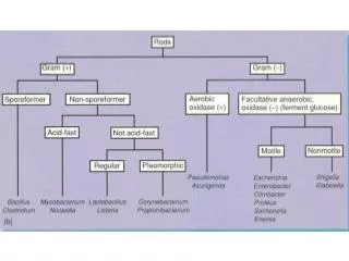
Click icon for audio Differentiation ofMajor GPR Genera • Gram stain • Morphology • Arrangement • Formation of spores • Catalase reaction
Click icon for audio GPR Discussed in Student Lab • Spore-forming GPR, catalase positive • Bacillus species • Bacillus anthracis • Bacillus cereus • Nonspore-forming GPR, catalase positive • Listeria monocytogenes • Corynebacterium species (diphtheroids) • Corynebacterium diphtheriae • Corynebacterium jekeium
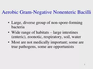
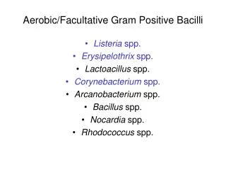
Click icon for audio GPR Discussed in This Lecture • Nonspore-forming GPR, catalase negative • Gardnerella species • Erysipelothrix species • Lactobacillus species • Branching Nocardioform GPR • Nocardia species • Streptomyces species
GPR, pleomorphic, Non-spore forming, Catalase negative Click icon for audio Gardnerella vaginalis Gram Stain • Pleomorphic gram-variable bacilli • Non-sporulating • Specimen gram stain: • Clue cells
Does not grow on sheep blood agar Human Blood Agar (V-agar, HBT-agar) Small colonies with diffuse zone of beta-hemolysis GPR, pleomorphic, Non-spore forming, Catalase negative Click icon for audio Gardnerella vaginalisColony Morphology
GPR, pleomorphic, Non-spore forming, Catalase negative Click icon for audio Gardnerella vaginalisIdentification • Catalase negative • Oxidase negative • Sodium hippurate usually positive • SPS sensitive
GPR, pleomorphic, Non-spore forming, Catalase negative Click icon for audio Gardnerella vaginalisClinical Significance • Normal vaginal flora • Bacterial vaginosis • Polymicrobic infection with Mobiluncus and Bacteroides • Diagnosis • Homogeneous, gray discharge • Clue cells • Amine or fishy odor when add 10% KOH • Culture not necessary for diagnosis
What are the key identification characteristics to determine if Gardnerella vaginalis is the cause of bacterial vaginosis? Examine vaginal specimen for presence of a homogeneous, gray discharge, clue cells on the Gram stain, and an amine or fishy odor when adding 10% KOH. A culture is not recommended, but if done, G. vaginalis grows on V-agar/HBT-agar and it beta hemolytic.
GPR, pleomorphic, Non-spore forming, Catalase negative Click icon for audio Erysipelothrix rhusiopathiae Gram Stain • Two types • Short GPR • Long, filamentous GPR • Non-sporulating
GPR, pleomorphic, Non-spore forming, Catalase negative Click icon for audio Erysipelothrix rhusiopathiae Colony Morphology • Grows on BAP • Nonhemolytic or alpha hemolytic • Microaerophilic
GPR, pleomorphic, Non-spore forming, Catalase negative Click icon for audio Erysipelothrix rhusiopathiae Identification • Catalase negative • Motility • Nonmotile • Test-tube brush growth pattern in semisolid motility tube at 48 hours • H2S positive in KIA/TSI (only GPR) • Sucrose non“F”
GPR, pleomorphic, Non-spore forming, Catalase negative Click icon for audio Erysipelothrix rhusiopathiae Clinical Significance • Zoonotic • Swine erysipelas (fatal to pigs) • Man – skin disease • Erysipelas • Direct contact with infected animal • Rarely disseminates causing septicemia with arthritis or endocarditis
What are the key biochemical reactions that identify Erysipelothrix rhusiopathiae? Two gram stain morphologies (GPR), two colony types on SBA, catalase negative, H2S positive, test-tube brush pattern in motility tube, sucrose non“F”
GPR, pleomorphic, Non-spore forming, Catalase negative Click icon for audio Lactobacillus speciesGram Stain • Two types • Long slender GPR in chains • Short GPCB • Non-sporulating
GPR, pleomorphic, Non-spore forming, Catalase negative Click icon for audio Lactobacillus speciesColony Morphology • Grows on BAP • Multiple colony morphologies • Nonhemolytic or alpha hemolytic • Microaerophilic
GPR, pleomorphic, Non-spore forming, Catalase negative Click icon for audio Lactobacillus speciesIdentification • Catalase negative • Sucrose “F” • Vancomycin “R”
GPR, pleomorphic, Non-spore forming, Catalase negative Click icon for audio Lactobacillus speciesClinical Significance • Normal flora • Mouth • GI tract • Female vaginal tract • Rarely pathogenic • Endocarditis • Meningitis
What is the clinical significance of Lactobacillus species? It is considered normal flora of the mouth, gastrointestinal tract, and female genital tract
Click icon for audio Review • Nonspore-forming GPR, catalase negative • Gardnerella species • Erysipelothrix species • Lactobacillus species
Click icon for audio Branching Nocardioform GPR • Actinomycetes • Nocardia species • Streptomyces species
Branching Nocardioform GPR Click icon for audio Nocardia speciesGram Stain • Pleomorphic,branching, fine, delicate filaments with fragmentation GPR • Often appears beaded
Branching Nocardioform GPR Click icon for audio Nocardia speciesColony Morphology • Grows on SBA, Mycology media and LJ media • Aerobic growth appears at 3-30 days • Waxy, bumpy or velvety rugose forms, yellow to orange colonies
Branching Nocardioform GPR Click icon for audio Nocardia speciesIdentification • Partially acid-fast positive • Presence of granules in specimen • Catalasepositive
Branching Nocardioform GPR Click icon for audio Nocardia speciesClinical Significance • Habitat: soil and water • Mycetoma (actinomycetoma) • Tissue swelling • Draining sinus tracts • Presence of granules • Immunocompromised patients • Pulmonary and disseminated infections
What is the key characteristic in identifying Nocardia species? Branching GPR that is partially acid fast positive
What primary disease does Nocardia cause and what 3 symptoms are seen? Mycetoma (aka actinomycetoma). Triad of symptoms: tissue swelling, draining sinus tracts, and the presence of granules
Branching Nocardioform GPR Click icon for audio Streptomyces speciesGram Stain • GPR with extensive branching, chains and spores • Does not fragment easily
Branching Nocardioform GPR Click icon for audio Streptomyces speciesColony Morphology • Grows on SBA, Mycology media and LJ media • Aerobic growth appears at 3-30 days • Waxy, bumpy or velvety rugose forms, yellow to orange colonies
Branching Nocardioform GPR Click icon for audio Streptomyces speciesIdentification • Acid-fast negative
Branching Nocardioform GPR Click icon for audio Streptomyces speciesClinical Significance • Habitat: soil and decaying vegetation • Mycetoma (actinomycetoma) • Rarely: • Pericarditis • Bacteremia • Brain abscess
How can you differentiate Streptomyces from Nocardia? Streptomyces is acid-fast negative and Nocardia is partially acid-fast positive
Click icon for audio Aerobic GPRSummary • Nonspore-forming GPR, catalase negative • Gardnerella species • Erysipelothrix species • Lactobacillus species • Branching Nocardioform GPR • Nocardia species • Streptomyces species
Who am I? HBT media shows beta-hemolysis Gram Stain Causes Bacterial Vaginitis Gardnerella vaginalis
Who am I? BAP, growth at 2 days Gram Stain TSI Motility Tube: “test-tube brush” pattern of growth Erysipelothrix rhusiopathiae
Who am I? LJ agar at 1 week Acid Fast Stain Gram Stain Causes Actinomycetoma Nocardia species