Download
1 / 23
250 likes | 265 Views
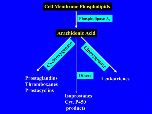
Nanoencapsulation Systems Based on Milk Proteins and Phospholipids. 식품공학실험실 2018246013 박지원. Introduction. What is Nanoencapsulation?. Techniques for wrapping encapsulated particulate functional material with a coating material.

E N D
Nanoencapsulation Systems Based on Milk Proteins and Phospholipids 식품공학실험실 2018246013 박지원
Introduction What is Nanoencapsulation? • Techniques for wrapping encapsulated particulate functional material with a coating material. • Depending on size, they are classified into macro, micro, and nano capsules.
Introduction What is Nanoencapsulation? • microencapsulation can be used stabilizing the core material, controlling the oxidative reaction, masking flavors, colors or odors, to extend shelf life or protect components against nutritional loss.
Introduction What is Nanoencapsulation? • These nanoparticles are able to encapsulate and deliver the active compounds directly to appropriate sites, maintain their concentration at suitable levels for long periods of time, and prevent their premature degradation. • Research efforts are already being made to develop food-based delivery vehicles, such as protein-polysaccharide coacervates, multiple emulsions, liposomes and cochleates.
Introduction What is Milk Protein? • Normal bovine milk contains about 3.5% protein which can be separated into caseins and whey proteins. • Caseins can be fractionated into four distinct proteins, αs1- , αs2-, β- and κ-caseins; these represent approximately 38%, 10%, 36% and 12% of whole caseins, respectively. • The principal fractions of whey proteins are β-lactoglobulin, bovine serum albumin, α-lactalbumin and immunoglobulins. In contrast to caseins, the whey proteins possess high levels of secondary, tertiary and, in most cases, quarternary structures.
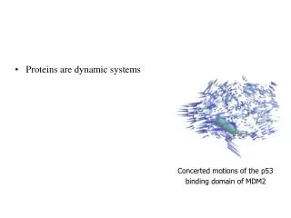
Introduction 1. Casein • The structures of caseins are quite unique. • → The most unusual feature is the amphiphilicity of their primary structure. • The hydrophobic residues and many of the charged residues, particularly the phosphoserine residues, in the caseins are not uniformly distributed along the polypeptide chain. • As an example, the distribution of charged residues and hydrophobicity as a function of sequence position of β-casein
Introduction 1. Casein
Introduction 2. Whey Protein • β-Lg is built up of two β-sheets, formed from nine strands converging at one end to form a hydrophobic calyx or pocket, and a flanking three-turn α-helix • → This pocket serves as a binding locus for apolar molecules such as • retinol and long-chain fatty acids. • Similarly, bovine serum albumin binds a large variety of compounds, including retinol and long-chain fatty acids.
Milk Proteins as Potential Nano-encapsulation Systems 1. Casein • Casein and caseinates can bind water, stabilise foams, emulsify fat and control viscosity in formulated foods, in addition to providing high nutritional value. • Caseins also possess many of the properties required of a good wall material for encapsulation. • A recent study showed that hydrophobic compounds, such as vitamin D2, can be incorporated into casein particles, formed by the re-assembly of caseins. These reassembled casein particles can provide partial protection against UV-light-induced degradation of vitamin D2 entrapped in them.
Milk Proteins as Potential Nano-encapsulation Systems 1. Casein
Milk Proteins as Potential Nano-encapsulation Systems 2. Whey Protein • The ability of whey proteins to aggregate and form gels during heat treatment is of considerable importance. • By controlling the assembly of protein molecules during the aggregation process, hydrogels, micro- and nanoparticles suitable for the delivery of bioactive compounds can be produced.
Milk Proteins-Polysaccharide Composite Systems • Protein structure can be modified through processing treatments or change of solution conditions to allow formation of complexes with polysaccharides. • At pH values below their isoelectric points (pI), proteins carry positive charges and can interact with polysaccharides bearing carboxylic, phosphate, or sulfate groups. This inter-biopolymer complexation of positively charge proteins and anionic polysaccharides can lead to the formation of soluble and insoluble complexes.
Milk Proteins-Polysaccharide Composite Systems • Recently, we have observed the formation of soluble and stable complexes on the mixing of gum arabic and sodium caseinate at a wide pH range (pH 4 to 5.4); the complexes resulted in stable dispersions with particle size between 100 to 200 nm.
Milk Proteins-Polysaccharide Composite Systems • As the pH of the mixture decreases below pH 5.4, the caseinate molecules tend to undergo small-scale aggregation prior to large-scale aggregation and precipitation at pH values closer to their pi (pH 4.6). • In this case, the gum arabic molecules may attach to the outside of these small-scale aggregates in the early stages of aggregation through electrostatic interactions between negatively charged gum arabic and exposed positive patches on the surface of the caseinate aggregates • The presence of hydrophilic gum arabic molecules on the outside of the caseinate aggregate may be enough to sterically stabilise these nano-particles and consequently prevent self-aggregation.
Effect of NaCl and CaCl2 on the properties of nanoparticles • Different amounts of NaCI or CaCl2 were added to the stable nanoparticle dispersion of caseinate/gum arabic at pH 4.6. The changes in the absorbance and average particle size of mixtures, which were measured after storage for 24 h, are shown in Figure 3.
Effect of NaCl and CaCl2 on the properties of nanoparticles • Effect of NaCl This indicated that the precipitation was due to self-association of caseinate molecules, and the association of gum arabic with the casein aggregates was prevented by the added NaCI. This is consistent with our previous work that showed that the complexes can not be formed between the sodium caseinate and gum arabic in the presence of NaCI > 50 mM. At added NaCI < 50 mM, the size of stable complex particles increased with increasing the concentration of NaCI. This could be due to the dissociation of some of the gum arabic molecules from the casein aggregate surface.
Effect of NaCl and CaCl2 on the properties of nanoparticles • Effect of CaCl2 The behaviour of dispersions after addition of CaCl2 was similar to that after addition of NaCI. This indicated that the effect of added CaCl2 was because of change in the ionic strength of system. In addition, It was ion bridging that could take place between the divalent calcium ion and the negatively charged casein molecules in the complexes. This may result in an increase in the size of complex particles and precipitation above certain CaCl2 concentration.
Milk Phospholipids as Potential Nanoencapsulation Systems • Milk contains 0.13-0.34 mg mg-1 protein phospholipids of which about 60% are associated with milk fat globule membrane (MFGM). • The composition of the MFGM phospholipid is very different from the commonly used soy- or egg-derived phospholipids. These differences would be expected to influence the selfassembly characteristics of phospholipids produced from the MFGM.
Formation of Liposomes from MGFM-phospholipids • Liposomes are spherical structures, with diameters ranging from 20 nm toseveral microns, formed through the self-assembly of amphiphilic molecules, usually phospholipids. They consist of one or more phospholipid bilayers enclosing an aqueous core. During the formation process, hydrophobic molecules can be incorporated in the lipid bilayers and hydrophilic molecules become entrapped in the aqueous core. • Liposomes are used by the pharmaceutical and cosmetic industries for theentrapment and controlled release of drugs or nutraceuticals, as model membranes or cells, and even for specialist techniques such as gene delivery.
Formation of Liposomes from MGFM-phospholipids • We have recently shown that liposomes can be produced from milk-derived phospholipids, using a microfluidization technique (18-20). Typical electron micrograph of a liposome dispersion prepared using MFGM by this technique is shown in Figure 4.
Formation of Liposomes from MGFM-phospholipids • The liposomes prepared from the milk phospholipid material have been shown to have a significantly higher phase transition temperature, thicker membrane and lower membrane permeability than liposomes prepared from a non-hydrogenated soy phospholipid fraction. • Experiments comparing the stability of the liposome dispersions found that the milk phospholipid liposome dispersions showed significantly better stability during storage at temperatures in the range 4-35 °C. • The milk phospholipid dispersions were also more stable during a wide range of heat treatments and ionic environments.
Conclusion • Milk contains several components that can be utilized to make nanoparticles for encapsulation and delivery of bioactive compounds. • The ability of milk proteins to interact strongly with charged polysaccharides opens up further possibilities for making novel hybrid nanoparticles. • We are currently determining the potential for use of such liposomes for the delivery of bioactive material in food systems in terms of encapsulation and release of hydrophobic and hydrophilic materials within the liposomes.