Download
1 / 36
360 likes | 718 Views
Comparative effect of Various HDAC-inhibitors in-vitro on T-Cell Lymphoma cell lines alone and in combination with conventional anti-cancer drugs. Arshad H. Banday Mentor:Dr . Francisco Hernandez- Illizaliturri. Introduction.

E N D
Comparative effect of Various HDAC-inhibitors in-vitro on T-Cell Lymphoma cell lines alone and in combination with conventional anti-cancer drugs Arshad H. Banday Mentor:Dr. Francisco Hernandez-Illizaliturri
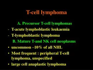
Introduction. • T-cell lymphomas are an uncommon and heterogeneous group of non-Hodgkin lymphomas. • Historically therapies for these diseases have been borrowed from treatments for other lymphomas. • More recently, efforts have be made to identify novel agents for their activity specifically in T-cell lymphomas. • A primary example of new agents with specific activity in T-cell lymphomas is the novel class of drug, histonedeacetylase inhibitors • Vorinostat and romidepsin are currently approved and are in clinical use for the treatment of cutaneous T-cell lymphomas.
Intro….. • Histones are core structural components of chromatin; DNA is wound around histones, and histones further associate to become and form chromatin. • Histone deacetylation inhibitors (HDAC) inhibitors induce accumulation of acetylated histones which leads to relaxation of chromatin structure and promotes access to transcriptional machinery and RNA polymerase • HDACi also modify other cancer related proteins.
Chromatin Structure Regulates Transcriptional Activity • HistoneDeacetylase Inhibitors (HDAC Inhibitors) • Cause increased histoneacetylation resulting in.. • Uncoiling of chromatin and transcriptional activation of tumor suppressor genes leading to cell cycle arrest and/or apoptosis • Currently only Vorinostat is licensed for use in cutaneous T cell lymphoma (CTCL)
EPIGENETIC Chromatin Enzyme modification errors Replication errors Mutations/translocations Open/closed chromatin DNA sequencenot altered DNA sequence altered Altered mRNA/proteins Altered DNA/mRNA/proteins Transformed cells • Can be caused by: • Abnormal modifications to histone proteins • Abnormal DNA methylation Oncogenesis Genetic Variations and Epigenetic Changes Can Both Contribute to Oncogenesis GENETIC DNA
Deacetylation of Histones by HDAC Can Prevent Gene Expression Acetylation by histone acetyltransferases (HATs) allows transcription and gene expression HAT Transcription factors HISTONE ACETYLATION HISTONE DEACETYLATION Deacetylated Histone Closed chromatin Transcription factors cannot access DNA Acetylated Histone Open chromatin Transcription factors can access DNA HDAC Deacetylation by histone deacetylases (HDACs) can prevent transcription and gene expression Ac: acetyl group HDAC depicts a class I deacetylase
In Tumor Cells, Imbalanced HAT and HDAC Activity Can Result in Deregulated Gene Expression HAT Decreased HAT Activity IncreasedHDAC Activity HDAC HDAC TF HDAC Decreased Tumor Suppressor Gene Activity (p21, p27) Tumor Unchecked CellGrowth and Survival Ac: acetyl group TF: transcription factors HDAC depicts a class I deacetylase
HDAC HDAC HDAC HDAC Inhibition Restores Gene Expression in Tumor Cells DAC Inhibition Increases Acetylation of Histones HAT TF DAC Inhibitor HDACi shifts balance Increased Tumor Suppressor Gene Activity (p21, p27) Normalized Cell Ac: acetyl group TF: transcription factors HDAC depicts a class I deacetylase Cell-Cycle Arrest and Differentiation Growth arrest
Loss oftumorsuppressor function Downstream effects Tumorsuppressorgene activity Microtubule depolymerization/ aggresome formation VEGF Oncoproteins Cell-cycle arrest Cell motility and Invasion Cell proliferation and survival Tumor effects Angiogenesis Apoptosis Deacetylase (DAC) Activity on Proteins is Associated with Downstream Effects that Promote Oncogenesis Proteins modulated by DACs Histone p53 HIF-1a -tubulin HSP90
DAC Inhibitor Loss oftumorsuppressor function Microtubule depolymerization/ aggresome formation Downstream effects Tumorsuppressorgene activity VEGF Oncoproteins Cell-cycle arrest Cell motility and Invasion Cell proliferation and survival Tumor effects Angiogenesis Apoptosis Pan-DAC Inhibition Interferes with the Multiple Hallmarks of Cancer Proteins modulated by DACs p53 -tubulin HSP90 Histone HIF-1a
Pan-DAC Inhibition May Have Potential in Several Cancers 50% of Cancers DAC Inhibitor Hematologic & Solid Tumors Histone p53 DACs HSP90 -tubulin HIF-1a CML, Breast, Prostate, NSCLC Breast, Multiple Myeloma RCC, Melanoma
Cells used • Loucy cell line: Loucy, was established from the peripheral blood of a patient with T-cell acute lymphoblastic leukemia. • HH Cell line: Cutaneous T- cell lymphoma. • SUP-T1: T-cell acute lymphoblastic leukemia
HDACi • Entinostat • Vorinostat Currently approved CTCL • LBH589 • Doses: 100, 10, 1, 0.1, 0.01 and 0.001 in MM
Other Materials • RPMI • 96 well plates. • Centrifuge tubes. • Cell counting chamber. • High power Microscope. • Colorimeter to measure fluorescence. • Alamar blue cell viability reagents. • Multi-channel micro- pipettes
Method. • Incubate each cell line with increasing dose of various HDACi
METHOD….. • 100 Microliter of T-cell lines in each well . • 100 microliter of HDAC inhibitor of various concentration or placebo was added to each well. • Incubate for 48 hour. • 100 Microliter of Alamar blue was added after 48 hours of Incubation.
METHOD… • Plates were Incubated for 4-6 Hours with Alamar blue. • Cell viability was measured by measuring fluorescence in each well using colorimeter. • Fluorescence in each drugged well was compared with the placebo and the number was plotted for various concentration using SSPS software • SSPS software was used for analysis. • Student’s t-test used for statistical analysis
HH P<0.05
Combination of panobinostat with: • Bortezomib • Doxorubicin • Cisplatin. • Gemcitabine
Flow(Cell cycle studies) • Treated each cell line with three different HDACi for 24,48 and 74 hours.
RESULTS: • All the three HDACi Exhibited potent killing effect on T-cell lymphoma cells lines in vitro. • Panobinostat is most potent of the HADCi studied and difference in activity was highly significant. • Panobinostat demonstrated additive killing effect in combination with Bortezomab and Doxirubacin. The additive effect is most likely due to different MOA.
Conclusion • The newer HDACi Panobinostat exhibited potent killing effect as compared to Vorinostat which is currently approved in the treatment T-cell lymphoma. • Combination with other anti-cancer drugs produced additive effect and holds promise for future. • The results would need to be validated by different method of assessing the killing effect and eventually testing on patient samples.
References • Li JY, Horwitz S, Moskowitz A, Myskowski PL, Pulitzer M, Querfeld C: Management of cutaneous T cell lymphoma: cancer management and research 2012,4:75-89 • Copeland A, Buglio D and Yones A; Histone deacetylseinhibitors in Lymphoma: Current opinion in oncology 2010, 22:431-436 • Horwitz SM: The Emerging Role of Histone Deacetylase Inhibitors in Treating T-cell lymphoma:CurrHematolMalig Rep 2011,1:67-72. • Garber K. HDAC inhibitors overcome first hurdle. Nat Biotechnol 2007;25:17–9. • Esteller M. Nat Rev Genet 2007;8:286–98 • Mehnert JM, Kelly K. Histone Deacetylase Inhibitors: Biology and mechanism of Action. Cancer J 2007;13:23–9. • MinucciS, Pelicci PG. Histone Deacetylase inhibitors and the promise of epigenetic (and more) treatments for cancer. Nat Rev Cancer 2006;6:38–51. • YooCB, Jones PA. Nat Rev Drug Discov 2006;5:37–50 • Kim DH, kim M, Kwon HJ Histone Deacetylase in Carcinogenesis and its Inhibitors as Anti-cancer Agent: Jour of Biochem and Mol Bio 2003,36 110-119 • Marks PA, Rifkind RA, Richon VM, Breslow R, Miller T and Kelly WK: Nat rev cancer 2001,1:194-202
Acknowledgement • Dr. Francisco Hernandez-Illizaliturri. • Dr. Myron Czuczman. • Dr. Khalid J Qazi. • Dr. Irfan Khan.