Download
1 / 28
280 likes | 449 Views
Oral Cavity. Lecture 23 Thursday, March 1, 2007 Refs. Moore and Dalley Chapter 2, Moore and Agur Chapter 8, Snell Chapter 5, Netter Atlas of Human Anatomy, Wheater’s Chapter 13, and Ross Chapter 16. Tooth composition and structure. Dentin forms the bulk of the tooth

E N D
Oral Cavity Lecture 23 Thursday, March 1, 2007 Refs. Moore and Dalley Chapter 2, Moore and Agur Chapter 8, Snell Chapter 5, Netter Atlas of Human Anatomy, Wheater’s Chapter 13, and Ross Chapter 16
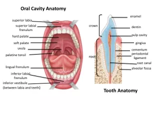
Tooth composition and structure • Dentin forms the bulk of the tooth • Calcified organic matrix similar to bone but harder (matrix has a greater proportion of inorganic material that is mainly in form of hydroxyapatite crystals). • Dentine tubules radiate from pulp cavity to periphery. • Enamel is an extremely hard translucent material • Parallel enamel rods or prisms cemented together by interprismatic material • Cementum is an amorphous calcified material. • Molars • Have >1 root • Have >1 cusp- sharp point on occlusal surface
Incisor (left) and molar (right)undecalcified ground sections 5 x WFH 13.4
Teeth have 2 embryological sources • Ectoderm- enamel • Epithelial ridge from the basal layer of oral epithelium forms a dental lamina that will become enamel organs • Mesoderm- dentin, cementum, pulp, and periodontal ligament • Mesenchyme beneath enamel organ becomes dental papilla • Interaction between ectoderm and mesoderm is important
Tooth formation at ~6weeks fetal (left) and after enamel organ has outlined shape of tooth crown (right). WFH 13.5a and c
Formation of dentin and enamel • Ameloblasts are cells that produce enamel. • Mineralized enamel contains less than 1% organic material and is the hardest and most dense substance in the body. • Enamel is incapable of regeneration. • Odontoblasts are cells that produce dentin. • Dentin is similar to bone but more mineralized • Deposition of dentin stimulates production of enamel. • Odontoblastic processes remain embedded in the matrix. • Root formation is not complete until after eruption.
Dentin tubules contain processes of odontoblasts. P=predentin (not mineralized yet) WFH 13.6b
Periodontal membrane • Cementocytes secrete cementum (calcified organic matrix similar to bone but mostly acellular). • Sharpey’s fibers slant from bone surface toward apex of root. • Suspension of tooth in sling allows cushioning. • Osteoclast resorption and osteoblast deposition on opposite side of alveolus allows movement in orthodontic treatment.
Gingiva • Attached gingiva covers alveolar bone. • Free gingiva surrounds tooth. • Gingival crevice- potential space extends to the cemento-enamel junction. • Gingivitis- inflammation of the gingiva • Gingival recession- exposes dentin • Sensitive to temperature and chemicals • Periodontal disease- disease of supporting structures of teeth
Gingival attachment WFH 13.9Enamel does not show in demineralized sections.Crevicular epithelium is thin and often the site of entry of pathogens.
Dentition • Primary (Deciduous) • 20 teeth • Per quadrant- 2 incisors, 1 canine, 2 molars • Begin forming at 6 weeks gestation. • Erupt from 6-30 months after birth. • Permanent • Begin replacing deciduous teeth at ~ 6 years. • 32 teeth • Per quadrant - 2 incisors, 1 canine, 2 premolars, 3 molars • The 3 molars (6 year, 12 year, and wisdom ~17-21 years) have no deciduous precursors.
Nerve and blood supply to teeth • Inferior alveolar nerve is sensory to lower teeth (mandibular branch of trigeminal). • Superior alveolar nerves supplies upper teeth (maxillary branch of trigeminal). • Inferior alveolar artery, a branch of maxillary artery, supplies lower teeth. • Superior alveolar arteries supply upper teeth.
Salivary glands • 3 Major salivary glands • Parotid, submandibular, and sublingual glands • Secrete in response to parasympathetic stimulation • Physical, chemical and emotional stimuli • Numerous minor salivary glands • In oral mucosa, tongue, palate • Secrete continuously
Saliva • Hypotonic secretion containing mucus, enzymes, antibodies, and ions. • Lysozyme is antibacterial. Amylase begins digestion of starches. • Saliva helps prevent tooth decay. • Keeps mucous membranes moist, aids ability to taste, and lubricates food during mastication and swallowing.
Submandibularmixed serous and mucous acini, often serous demilunes cap mucous acini WFH 13.17
Duct system of salivary glands • Intercalated duct • smallest, lined by cuboidal secretory cells • Striated duct • Tall columnar cells. Striations are due to interdigitations of basal cytoplasmic processes of adjacent cells. (mitochondria pack basal processes) • Reabsorb ions (Na+ and Cl-) from the isotonic secretion produced by serous cells and secrete K+ and HCO3- • Excretory duct • Stratified cuboidal
Waldeyer’s ring • Lymphatic tissue surrounds the posterior openings of the oral and nasal cavities. • Lingual tonsil • Palatine tonsils • Tubal tonsils in the lateral nasopharynx posterior to the opening of Eustachian tube • Pharyngeal tonsil (adenoid) in roof of nasopharynx