Download
1 / 69
690 likes | 866 Views
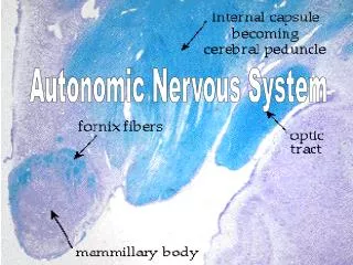
Chapter 12 Autonomic Nervous System. Autonomic or Visceral Reflexes. What They Do: Autonomic reflexes regulate organ function Pathway: The sequence is receptor activation, sensory input ( CNS), motor neuron response, and effector response.

E N D
Autonomic or Visceral Reflexes • What They Do: Autonomic reflexes regulate organ function • Pathway: The sequence is receptor activation, sensory input ( CNS), motor neuron response, and effector response
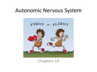
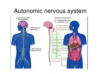
Organization and Function of the Autonomic Nervous System • Divisions of the ANS: There are two divisions. • Sympathetic nervous system, called “Fight or Flight.” • Parasympathetic Nervous System, called “Feed and Breed.” • Autonomic Terminology and Autonomic Pharmacology • Drugs that affect the sympathetic nervous system are called sympathomimetic and sympatholytic. • Drugs that affect the parasympathetic nervous system are called parasympathomimetic and parasympatholytic.
Organization and Function of the Autonomic Nervous System - cont’d • Autonomic Tone and Vasomotor Tone • Background firing of the ANS causes autonomic tone. • Background sympathetic stimulation of the blood vessels causes vasomotor tone.
ANS: Neurons • Numbers and Ganglia • Preganglionic fibers are fibers that extend from the CNS to the ganglia. • Postganglionic fibers are fibers that extend from the ganglia to the effector organ.
ANS: Neurons - cont’d • Neurons of the Sympathetic Nervous System • The SNS is called the thoracolumbar outflow. • The sympathetic ganglia are located in a chain close to the spinal cord; the chain is called paravertebral ganglia. • The adrenal medulla secretes hormones that mimic the SNS.
ANS: Neurons - cont’d • Neurons of the Parasympathetic Nervous System • The parasympathetic nervous system is called the craniosacral outflow. • Parasympathetic fibers travel with cranial nerves; most parasympathetics run with the vagus nerve CN X. • Naming Fibers and Neurotransmitters • Cholinergic fibers secrete acetylcholine (ACh). • Adrenergic fibers secrete norepinephrine (NE).
ANS: Neurons - cont’d • Neurotransmitters: Termination of Activity • ACh is degraded immediately by acetylcholinesterase. • NE activity is ended primarily by reuptake of the NE into the nerve terminal and by MAO activity within the nerve terminal.
Receptors of the Autonomic Nervous System • Cholinergic Receptors • These are activated by ACh. • There are two types: muscarinic and nicotinic (with subtypes). • Adrenergic Receptors • Activated by NE • There are two types: alpha and beta (with subtypes).
Receptors of the Autonomic Nervous System - cont’d • Receptor activation and blockade can be determined by examining Tables 12-1, 12-3, and 12-4. • Autonomic Receptors: “Doing Autonomic Pharmacology” • Clinical examples where drugs target autonomic receptors
Introduction • The brain, spinal cord, and peripheral nervous system act as a vast communication system. The spinal cord transmits information to and from the brain. The peripheral nervous system brings information to the CNS (its sensory role) and delivers information from the CNS to the periphery (its motor role).
What the Spinal Cord Is • The spinal cord is a tubelike structure located in the spinal cavity, extending from the foramen magnum (occipital bone) to L1 • Arrangement of Nervous Tissue • The gray matter is a centrally located, butterfly-shaped area. • The white matter is composed of myelinated fibers arranged in tracts. Ascending tracts are sensory tracts. Descending tracts are motor tracts.
What the Spinal Cord Is - cont’d • Arrangement of Nervous Tissue—cont’d • Spinal nerves are attached to the spinal cord. All spinal nerves are mixed (they contain sensory and motor fibers). • Sensory nerve fibers travel to the cord through the dorsal root. Motor nerve fibers travel in the ventral root.
What the Spinal Cord Does: Functions • The spinal cord relays both sensory and motor information. • The spinal cord acts as a major reflex center.
Reflexes • A reflex is an involuntary response to a stimulus. • The four components to a reflex are a sensory receptor; an afferent (sensory) neuron; an efferent (motor) neuron; and an effector organ.
Peripheral Nervous System • Nerve • A nerve is a group of neurons, blood vessels, and connective tissue. • There are sensory nerves, motor nerves, and mixed nerves.
Peripheral Nervous System - cont’d • Structural Classification of Nerves • A classification of nerves on the basis of structure divides nerves into cranial nerves and spinal nerves. There are 12 pairs of cranial nerves (Table 11-3) and 31 pairs of spinal nerves (Table 11-5). • Spinal nerves are sorted out at nerve plexuses. The three major plexuses are the cervical plexus, the brachial plexus, and the lumbosacral plexus. • A dermatome is the area of skin innervated by each spinal nerve.
Peripheral Nervous System - cont’d • Functional Classification of Nerves • Somatic afferent nerves carry sensory information to the CNS. • Somatic efferent nerves carry motor information to skeletal muscles. • Autonomic nerves carry motor information to the organs (viscera).
Introduction • The purpose of the nervous system is to bring information to the central nervous system, interpret the information, and enable the body to respond to the information.
The Nervous System: Overview • Divisions of the Nervous System • The central nervous system (CNS) includes the brain and the spinal cord. • The peripheral nervous system includes the nerves that connect the CNS with the rest of the body.
The Nervous System: Overview - cont’d • Cells That Make Up the Nervous System • Neuroglia (glia) support, protect, and nourish the neurons. • Neurons conduct the nerve impulse. • The three parts of a neuron are the dendrites, cell body, and axon.
The Nervous System: Overview - cont’d • Types of Neurons • Sensory, or afferent, neurons carry information toward the CNS. • Interneurons are located in the CNS (make connections). • Motor, or efferent, neurons carry information away from the CNS toward the periphery.
The Nervous System: Overview - cont’d • White Matter and Gray Matter • White matter is due to myelinated fibers. • Gray matter is composed primarily of cell bodies, interneurons, and unmyelinated fibers. • Clusters of cell bodies (gray matter) are called nuclei and ganglia.
The Neuron Carrying Information • Nerve Impulse • The electrical signal is called the action potential or nerve impulse. • The nerve impulse is due to the following changes in the neuron: polarization, depolarization, and repolarization. • The nerve impulse is due to flow of ions: polarization (outward flux of K+), depolarization (influx of Na+), and repolarization (outward flux of K+).
The Neuron Carrying Information - cont’d • Nerve Impulse—cont’d • The refractory period is the unresponsive period of the neuron. • The nerve impulse jumps from node to node as it travels along a myelinated fiber. Myelination increases the speed of the nerve impulse. • The nerve impulse causes the release of a neurotransmitter.
The Neuron Carrying Information - cont’d • Synapse • The synapse is a space between two neurons. • The nerve impulse of the first (presynaptic) neuron causes the release of neurotransmitter into the synaptic cleft. The neurotransmitter diffuses across the synaptic cleft and binds to the receptors on the second (postsynaptic) membrane. The activation of the receptors stimulates a nerve impulse in the second neuron.
Brain: Structure and Function • Cerebrum • The right and left hemispheres are joined by the corpus callosum. • The four main cerebral lobes are the frontal, parietal, temporal, and occipital lobes. Functions of each lobe are summarized in Table 10-2. • Large areas of the cerebrum, called association areas, are concerned with interpreting, integrating, and analyzing information.
Brain: Structure and Function - cont’d • Diencephalon • The thalamus is a relay station for most sensory tracts traveling to the cerebrum. • The hypothalamus controls many body functions such as water balance, temperature, and the secretion of hormones from the pituitary gland; it exerts an effect on the autonomic nervous system.
Brain: Structure and Function - cont’d • Brain Stem • Brain stem: midbrain, pons, and medulla oblongata. • The medulla oblongata is called the vital center because it controls the heart rate, blood pressure, and respirations (the vital functions). • The vomiting center is located in the medulla oblongata; it receives input directly and indirectly from activation of the chemoreceptor trigger zone (CTZ).
Brain: Structure and Function - cont’d • Cerebellum • The cerebellum is sometimes called the little brain. • The cerebellum is concerned primarily with the coordination of voluntary muscle activity.
Brain: Structure and Function - cont’d • Structures Involving More than One Lobe • The limbic system is sometimes called the emotional brain. • The reticular formation is concerned with the sleep/wake cycle. It keeps us conscious and prevents us from slipping into a coma state. • The “memory areas” handle short-term and long-term memory.
Protection of the CNS • Bone: cranium and vertebral column • Meninges: pia mater, arachnoid, and dura mater • Cerebrospinal fluid (CSF) that circulates within the subarachnoid space • Blood-brain barrier
Introduction • The purpose of muscle is to contract and to cause movement.
Muscle Function: Overview • Types and Functions of Muscles • Skeletal muscle is striated and voluntary; its primary function is to produce movement. • Smooth (visceral) muscle is nonstriated and involuntary; it helps the organs perform their functions. • Cardiac muscle is striated and involuntary; it is found only in the heart and allows the heart to function as a pump.
Muscle Function: Overview - cont’d • Structure of the Whole Muscle • A large muscle consists of thousands of single muscle fibers (muscle cells). • Connective tissue binds the muscle fibers (cells) together (forming compartments in the limbs) and attaches muscle to bone and other tissue (by tendons and aponeuroses).
Muscle Function: Overview - cont’d • Structure and Function of a Single Muscle Fiber • The muscle fiber (cell) is surrounded by a cell membrane (sarcolemma). The cell membrane penetrates to the interior of the muscle as the transverse tubule (T tubule). • An extensive sarcoplasmic reticulum (SR) stores calcium. • Each muscle fiber consists of a series of sarcomeres. Each sarcomere contains the contractile proteins actin and myosin.
Muscle Function: Overview - cont’d • How Muscles Contract • Muscles shorten or contract as the actin and myosin (in the presence of calcium and ATP) interact through crossbridge formation, according to the sliding filament theory. • For skeletal muscle to contract, it must be stimulated by a motor nerve. The nerve impulse releases acetycholine (ACh) from the nerve terminal. ACh diffuses across the neuromuscular junction (NMJ), binds to the muscle membrane and causes an electrical signal to form in the muscle membrane.
Muscle Function: Overview - cont’d • How Muscles Contract—cont’d • The electrical signal enters the T-tubular system and stimulates the SR to release calcium. • Actin, myosin, and ATP interact to form crossbridges, which cause sliding or shortening. • Calcium is pumped back into the SR and the muscles relax.
Muscle Function: Overview - cont’d • Responses of a Whole Muscle • A single muscle fiber contracts in an all-or-nothing response; a whole muscle can contract partially (i.e., not all-or-nothing). • A whole muscle increases its force of contraction by recruitment of additional muscle fibers. • Two terms describe the contractile activity of a whole muscle: twitch and tetanus. Tetanus refers to a sustained muscle contraction. • Energy for muscle contraction can be obtained from three sources: burning fuel aerobically, burning fuel anaerobically, and metabolizing creatine phosphate.
Muscle Function: Overview - cont’d • Terms That Describe Muscle Movement • Origin and Insertion: The attachments of the muscles. • Prime mover: The muscle most responsible for the movement achieved by the muscle group • Synergist and Antagonist: Works with, or has an opposing action.
Muscles from Head to Toe • Skeletal muscles are named according to size, shape, direction of fibers, location, number of origins, place of origin and insertion, and muscle action. • See Table 9-1 for a list of the body’s muscles.
Introduction • The skeletal system supports the weight of the body, supports and protects body organs, enables the body to move, acts as storage site for minerals, and produces blood cells.
Bones: An Overview • Sizes and Shapes • Bones are classified as long, short, flat, and irregular. • Bone markings function as sites of muscle attachments and passages for nerves and blood vessels. • A long bone has a diaphysis (shaft) and two epiphyses (ends). Articular cartilage is found on the outer surface of the epiphyses. • The diaphysis is composed of compact or hard bone. The epiphysis consists of spongy or soft bone; red marrow is found in the holes of spongy bone.
Bones: An Overview - cont’d • Bone Formation and Growth • Bones ossify in two ways. In the skull, osteoblasts replace thin connective tissue membrane, forming flat bones. Other bones form on hyaline cartilage models as osteoblasts replace cartilage with bone. • Bones grow longitudinally at the epiphyseal disc, to determine height; bones also grow thicker and wider to support the weight of the body. • Bone growth and reshaping occur throughout life and depend on many factors, including diet, exercise, and hormones.
Divisions of the Skeletal System • The names of the 206 bones of the skeleton are listed in Table 8-2.
Divisions of the Skeletal System - cont’d • Axial Skeleton • The axial skeleton includes the bones of the skull (cranium and face), hyoid bone, bones of the middle ear, bones of the vertebral column, and the thoracic cage. • The skull of a newborn contains fontanels, which are membranous areas that allow brain growth. • The skull contains air-filled cavities called sinuses.
Divisions of the Skeletal System - cont’d • Axial Skeleton—cont’d • The vertebral column is formed from 26 vertebrae, one sacrum, and one coccyx. The vertebrae are separated by cartilaginous discs. The vertebral column of the adult has four curvatures: cervical, thoracic, lumbar, and sacral. • The thoracic cage is a bony, cone-shaped cage formed by the sternum, 12 pairs of ribs, and thoracic vertebrae.
Divisions of the Skeletal System - cont’d • Appendicular Skeleton • The appendicular skeleton includes the bones of the extremities (arms and legs), and the bones of the hip and shoulder girdles. • The shoulder girdle consists of the scapula and the clavicle. • The pelvic girdle is formed by the two coxal bones and is secured to the axial skeleton at the sacrum.
Joints • A joint or articulation is the site where two bones meet.
Joints - cont’d • Types of Joints (based on the degree of movement) • Immovable joints. • Slightly movable joints. • Freely movable joints or synovial joints. Structures within a synovial joint (knee): articular cartilage, the joint capsule, synovial membrane, synovial fluid, bursae, and supporting ligaments. • The types of freely movable joints include hinge, ball and socket, pivot, gliding, saddle, and condyloid.