Download
1 / 1
10 likes | 134 Views
Problem. What effect will a site-specific mutation to the Pin1 protein have on catalytic efficiency? . Hypothesis. The mutations I28A and C113A will have different catalytic rates compared to regular, wild type Pin1 protein. Materials. LiCl Tetrahydrofuran (THF) Suc-AEPF-pNA HEPES

E N D
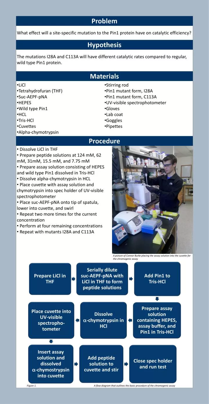
Problem What effect will a site-specific mutation to the Pin1 protein have on catalytic efficiency? Hypothesis The mutations I28A and C113A will have different catalytic rates compared to regular, wild type Pin1 protein. Materials • LiCl • Tetrahydrofuran (THF) • Suc-AEPF-pNA • HEPES • Wild type Pin1 • HCL • Tris-HCl • Cuvettes • Alpha-chymotrypsin • Stirring rod • Pin1 mutant form, I28A • Pin1 mutant form, C113A • UV-visible spectrophotometer • Gloves • Lab coat • Goggles • Pipettes Procedure • Dissolve LiCl in THF • Prepare peptide solutions at 124 mM, 62 mM, 31mM, 15.5 mM, and 7.75 mM • Prepare assay solution consisting ofHEPES and wild type Pin1 dissolved in Tris-HCl • Dissolve alpha-chymotrypsin in HCL • Place cuvette with assay solution and chymotrypsin into spec holder of UV-visible spectrophotometer • Place suc-AEPF-pNA onto tip of spatula, lower into cuvette, and swirl • Repeat two more times for the current concentration • Perform at four remaining concentrations • Repeat with mutants I28A and C113A A picture of Connor Burke placing the assay solution into the cuvette for the chromogenic assay Figure 1. A flow diagram that outlines the basic procedure of the chromogenicassay