Download
1 / 13
200 likes | 1.05k Views
Gross Anatomy of Skeletal Muscle. Connective tissue wrapping endomysium - fine sheath of areolar connective tissue wrapping each muscle fiber perimysium - collagenic sheath wrapping several muscle fibers gathered side by side into bundles, or fascicles

E N D
Gross Anatomy of Skeletal Muscle • Connective tissue wrapping • endomysium- fine sheath of areolar connective tissue wrapping each muscle fiber • perimysium- collagenic sheath wrapping several muscle fibers gathered side by side into bundles, or fascicles • epimysium- dense fibrous sheath wrapping several fascicles; surrounds entire muscle • deep fascia- coarse sheet of fibrous tissue that binds muscles into functional groups; extends to wrap other structures
Gross Anatomy of Skeletal Muscle • All of these connective tissue sheaths are continuous with one another and with the tendons; transmit force of muscle fibers to bone to be moved
Gross Anatomy of Skeletal Muscle • Nerve and Blood Supply a. each muscle fiber is supplied with a nerve ending that controls its activity b. muscles need a continuos supply of oxygen and nutrients by way of an artery; each muscle is served by one artery c. each muscle is served by one or more veins to remove large amounts of metabolic wastes produced during contraction
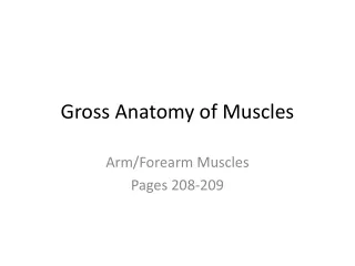
Gross Anatomy of Skeletal Muscles • Attachments • origin- the attachment of a muscle to an immovable bone • insertion- the attachment of a muscles to a movable bone • direct attachment- the epimysium of muscle is fused to the periosteum of bone or the perichondrium of cartilage • indirect attachment- the connective tissue wrappings extend beyond the muscle as a ropelike tendon or a flat, broad aponeurosis; more common and smaller in size
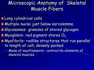
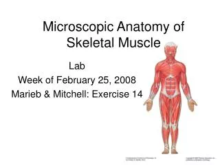
Microscopic Anatomy of Skeletal Muscle • Sarcolemma- plasma membrane surface of muscle fibers/cells • Sarcoplasm- cytoplasm of muscle cells; contains large amounts of stored glycogen and an oxygen-binding protein, myoglobin
Microscopic Anatomy of Skeletal Muscle • Myofibrils- rodlike, contractile elements extending the entire length of the cell; hundreds to thousands in each fiber/cell (80% of cell volume)
Microscopic Anatomy of Skeletal Muscle • striations- repeating series of dark and light bands along myofibril • A bands- dark bands • H zone- lighter stripe in midsection of an A band; visible when muscle is relaxed • M line- dark line bisecting each H zone ii. I bands- light bands - Z line- dark line bisecting each I band
Microscopic Anatomy of Skeletal Muscle • Sarcomere: region of myofibril between two successive Z lines; smallest contractile unit of a muscle fiber (Myofibrils are chains of sarcomeres aligned end-to-end)
Microscopic Anatomy of Skeletal Muscle • Myofilaments i. thick filaments- extend entire length of A band; composed of bundles of the protein myosin (rodlike tail, two globular heads) - cross bridges- myosin heads; link thick and thin filaments together during contraction ii. thin filaments- extend across the I band and partway into the A bands; composed of chains of globular protein actin - two strands of tropomysin spiral around actin chains troponin complex
Microscopic Anatomy of Skeletal System • Sarcoplasmic reticulum- elaborate smooth E.R. surrounding each myofibril; regulates levels of calcium ions (releases it on demand when muscle fiber is stimulated to contract)