Download
1 / 49
541 likes | 1.01k Views
Chapter 6 Bones and Skeletal Tissues. Part B Bone Structure. Bones. Bones are organs! Contains various types of tissues Osseous tissue (dominates) Nervous tissue Cartilage Fibrous connective tissue (lining cavities) Muscle and epithelial tissues in blood vessels. Bones.

E N D
Chapter 6Bones and Skeletal Tissues Part B Bone Structure
Bones • Bones are organs! • Contains various types of tissues • Osseous tissue (dominates) • Nervous tissue • Cartilage • Fibrous connective tissue (lining cavities) • Muscle and epithelial tissues in blood vessels
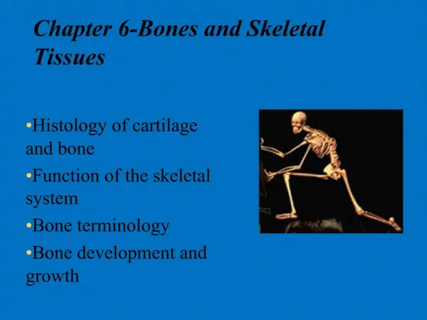
Bones • We will look at bone structure on three levels • Gross • Microscopic • Chemical
Bone Structure Gross Anatomy Bone Markings, Bone Textures, Bone Structures
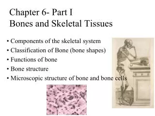
Bone Markings • Surface features of bones • Sites for attachments for muscles, tendons and ligaments • Joint surfaces • Passages for nerves and blood vessels
Bone Markings • Categories of bone markings • Projections • Bulges that grow outward from the bone surface • Depressions and openings • Indentations, holes, and cavities
ProjectionsThat Are Sites of Muscle and Ligament Attachment • Tuberosity – rounded projection • Crest – narrow, prominent ridge of bone • Trochanter – large, blunt, irregular surface • Line – narrow ridge of bone • Tubercle – small rounded projection • Epicondyle – raised area above a condyle • Spine – sharp, slender projection • Process – any bony prominence
Projections That help to Form Joints • Head – bony expansion carried on a narrow neck • Facet – smooth, nearly flat articular surface • Condyle– rounded articular projection • Ramus – armlike bar of bone
Depressions and OpeningsAllowing Blood Vessels & Nerves to Pass • Meatus – canal-like passageway • Sinus – cavity within a bone • Fossa – shallow, basinlike depression • Groove – furrow • Fissure – narrow, slitlike opening • Foramen – round or oval opening through a bone
Bone Textures • Compact bone – dense outer layer • Looks solid to the eye • Spongy bone – honeycomb or spongy appearance • Has small needle-like or flat pieces called trabeculae • filled with red or yellow bone marrow
Structure of Long Bone • General structure of Long Bones • Diaphysis • Epiphyses • Membranes
Structure of Long Bone • Diaphysis • Shaft constructed of a thick collar of compact bone that surrounds the medullary cavity
Structure of Long Bone • Medullary Cavity • Cavity of the shaft • Contains yellow marrow (mostly fat) in adults • Contains red marrow (for blood cell formation) in infants
Structure of Long Bone • Epiphyses • Expanded ends of long bones • Exterior is compact bone, interior is spongy bone • Joint surface is covered with articular cartilage • Epiphyseal line separates the diaphysis from the epiphyses
Structure of Long Bone • Articular cartilage • Covers the external surface of the epiphyses • Made of hyaline cartilage • Decreases friction at joint surfaces
Structure of Long Bone • Epiphyseal Line • Remnant of epiphyseal plate • Disc of hyaline cartilage that grows during childhood to lengthen the bone
Structure of Long Bone • Membranes of long bones • Periosteum • Endosteum
Structure of Long Bone • Periosteum • Outside covering of the bone • Except joint surfaces • Glistening white, double-layered membrane
Structure of Long Bone • Periosteum • Outer Fibrous layer is dense connective tissue • Inner osteogenic layer (next to bone surface) is mostly osteoblasts (bone germinators) and osteoclasts (bone breakers)
Structure of Long Bone • Periosteum • Richly supplied with nerve fibers, lymphatic vessels, blood vessels which enter the diaphysis by a nutrient foramen (opening)
Structure of Long Bone • Sharpey’s fibers • Secures periosteum to underlying bone • Made of collagen fibers • Extends into bone matrix
Structure of Long Bone • Arteries • Supplies bone cells with nutrients
Structure of Long Bone • Endosteum • Delicate connective tissue • Contains osteoblasts & osteoclasts
Structure of Long Bone • Endosteum • Covers internal surfaces of bone • Medullary cavity • Trabeculae (spongy) • Canals through compact bone
Structure of Long Bone Figure 6.3
Structure of Short, Irregular, and Flat Bones • Thin plates of periosteum-covered compact bone on the outside with endosteum-covered diploë on the inside • Spongy bone in flat bone is called diploë
Structure of Short, Irregular, and Flat Bones • Have no diaphysis or epiphyses • Contain bone marrow between the trabeculae
Red Bone Marrow Hematopoietic Tissue
Location of Hematopoietic Tissue (Red Marrow) • In infants • Found in the medullary cavity and all areas of spongy bone
Location of Hematopoietic Tissue (Red Marrow) • In adults • Found in the diploë of flat bones, and the head of the femur and humerus • Remember, the diploë is the spongy bone of flat bones
Yellow Bone Marrow • Yellow marrow is found in the medullary cavities • Yellow marrow can revert back to red marrow if a person becomes very anemic and needs enhanced red blood cell production
Bone Structure Microscopic Anatomy
Microscopic Structure of Compact Bone • Haversian system, or osteon • the structural unit of compact bone
Microscopic Structure of Compact Bone • Each osteon is a group of hollow tubes of bone matrix, called lamella, each one placed outside the next
Microscopic Structure of Compact Bone • Each osteon is oriented parallel to the long axis of the bone
Microscopic Structure of Compact Bone • Osteons are like tiny weight-bearing pillars
Microscopic Structure of Compact Bone • Lamella • The weight-bearing, column—like matrix tubes composed mainly of collagen
Microscopic Structure of Compact Bone • Haversian, or central canal • channel in the center of the osteon • containing blood vessels and nerves
Microscopic Structure of Compact Bone • Volkmann’s canals • channels lying at right angles to the central canal, connecting blood and nerve supply of the periosteum to that of the central canal
Microscopic Structure of Bone: Compact Bone • Osteocytes • Mature bone cells • Spider-shaped • Lacunae • small cavities in bone that contain osteocytes
Microscopic Structure of Bone: Compact Bone • Canaliculi • Tiny, hair-like canals • connect lacunae to each other and the central canal • Forms a transport system
Microscopic Structure of Compact Bone Figure 6.6a, b
Chemical Composition of Bone Organic components and inorganic components
Chemical Composition of Bone: Organic • Osteoblasts – bone-forming cells • Osteocytes– mature bone cells • Osteoclasts – large cells that resorb or break down bone matrix • Osteoid– unmineralized bone matrix composed of proteoglycans, glycoproteins, and collagen
Chemical Composition of Bone: Inorganic • Hydroxyapatites, or mineral salts • Sixty-five percent of bone by mass • Mainly calcium phosphates • Responsible for bone hardness and its resistance to compression
Chemical Composition of Bone: Inorganic • Hydroxyapatites, or mineral salts • Bone is half as strong as steel in resisting compression and fully as strong as steel in resisting tension • Bones last long after death, sometimes many centuries
Study Guide Pages 123 to top of 126 Quiz Next Time!