Download
1 / 70
720 likes | 1.03k Views
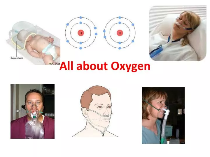
All about Oxygen. Introduction. As you know… It is tasteless, ordorless Supports combustion Vital for life May be mixed with other gases (He, CO2, N2, NO2) Comprises 21% of the atmopheric gas Normally 80-100 mmHg in the blood, 100 in the lung, 40 in the venous blood. Introduction.

E N D
Introduction • As you know… • It is tasteless, ordorless • Supports combustion • Vital for life • May be mixed with other gases (He, CO2, N2, NO2) • Comprises 21% of the atmopheric gas • Normally 80-100 mmHg in the blood, 100 in the lung, 40 in the venous blood
Introduction • As you know… • Critical temperature is 118.6 °C (-181 °F) • Can be stored as a liquid or gas • Produced through fractional distillation • Stored in cylinders or bulk supply systems • Used in the home with concentrators • Is a FDA drug, must be at least 99% pure, prescribed by a MD and approved by insurance before use by a pulmonary stress test
Introduction • As you know… • Filtered in a concentrator by a molecular sieve, gives up to 90% O2 • Semi permeable membranes give up to 40% O2 • May be blended to form Heliox • Given in either a high flow or low flow environment
Indications for Oxygen • Pneumothorax: Give 100% for Nitrogen washout • COPD/chronic lung disease: Low concentrations (below 30%) • Pulmonary Fibrosis: low flow, 25-40% O2 • Trauma/emergencies: typically give 100% O2 with a non-rebreathing mask • Ex: Car accident, hypovolemia
Indications for O2 • Dyspnea • Increased WOB/SOB • MI (up to 4L) • CVA, chest pain, high altitude sickness, pulmonary edema, restrictive diseases… • Orthopnea • Tachypnea • Pre-suctioning • Platypnea • Low Spo2, hypoxemia, anemia, hypoxia…. • If a patient is not breathing: Must supply positive pressure with 100% O2 via bag/mask
Indications for O2 • Without proper oxygenation a patient will develop hypoxemia which will produce symptoms of: • Tachypnea • Tachycardia • Cyanosis • Hypoxia/cell death • Anoxia/brain death • Heart failure • Low CaO2, PaO2, SaO2, SpO2 • Confusion, lethargy, weakness, malnutrition • SOB/WOB/Dyspnea…
Indications • Oxygen is used to: Decrease Ventilatory demand Decrease Work of breathing Decrease Cardiac output • Oxygen is required when: • SaO2 less than 90% • PaO2 less than 60 mm Hg
Determining the requirement of oxygen you must assess • Neurologic status (is there lethargy, anoxic events…) • Pulmonary status (check all O2 indices and assessments) • Cardiac status (HR, recent MI, poor EF, cardiac inflammation, valve problems…)
Why is Oxygen considered toxic? • High inspired oxygen concentrations cause toxicity by causing formation of oxygen free radicals (which damage tissues), and by causing absorption atelectasis and V/Q mismatch. • The issue of oxygen toxicity has been topical for a generation, following the discovery that therapeutic oxygen causes blindness in premature babies (retrolental fibroplasias) with respiratory distress syndrome. In addition, it has been established that high inspired concentrations of oxygen may cause acute lung injury, probably due to oxygen free radical production – superoxide, hydroxyl, hydrogen peroxide and singlet O2 molecules.
Why is Oxygen considered toxic? • These agents damage biomolecules such as membrane lipids, enzymes and nucleic acids. The extent of injury appears to depend on: • 1. The FiO2, • 2. The duration of exposure, • 3. The barometric pressure under which exposure occurred. • It appears that the critical FiO2 for toxicity is around 50% (1), above which lung recruitment maneuvers should be condidered (CPAP). • High concentrations of inspired oxygen may cause absorption atelectasis. In addition high FiO2 may cause increased peripheral vascular resistance in congestive heart failure leading to reduced cardiac output.
Why is Oxygen considered toxic? • How much oxygen is safe is a moot point. It is more important that you do not withhold life saving oxygen therapy than to be concerned about oxygen toxicity. It is, nonetheless, important that FiO2 is minimized to normalization of blood gas in intensive care patients: i.e. there is little to be gained in having a PaO2 of greater than 100mmHg. Often elevated oxygen requirements can be compensated for by appropriate patient positioning and increasing mean airway pressures to improve matching of ventilation and perfusion.
Oxygen Toxicity • Oxygen can be toxic, with high concentrations (>50%) over 24 hours it can lead to: · Decreased DLCO · Decreased CL · Increase PAO2 – PaO2 gradient · Decreased VC · Bilateral infiltrates on CXR · Absorption atelectasis · O2 induced hypoventilation · Formation of hyaline membrane · ROP
Titrate O2!! • Wean patients off O2 or to a safe FIO2 as soon as tolerated in order to avoid complications associated with O2 toxicity • Acceptable SpO2 Ranges: • COPD/Pulmonary Fibrosis: 88-92% • Adults w/o Lung disease: >92% • Pediatrics: >94% • Recent MI: >94%
Wean O2 • Discontinue O2 once the SpO2 reaches 92% on adult patients to avoid toxicity, wean FIO2 if SpO2 is greater than 92% • Oxygen can be administered by low flow or high flow devices. High flow devices include venturi masks which can be affected by: • Blocked entrainment ports (causes the devices total output flow to decreases, increases the percent of O2 from less entrained room air and it also changes the FIO2 received by the patient)
If a patient develops SOB on a venturi mask, assure that total flow is adequate, increase flow to mask as needed • Venturimasks go up to 50% FIO2 • Stable FIO2 requires the mask to provide all the gas needed by the patient during inspiration. • They can provide variable FIO2 values under some clinical conditions. • They always deliver O2 concentrations less than 100%. • yield a set FIO2 only if their flow exceeds the patient's.
High Flow Oxygen • Actual O2 provided by an air-entrainment system depends on: • O2 input flow to the jet • air-to-O2 ratio of the device • resistance downstream from the jet • Most low flow oxygen devices are not stable, they are easy to apply, disposable, have a low cost but are not stable, meaning they can come off the patients face easily.
Assessing O2 • Remember oxygenation is reflected by looking at PaO2 while ventilation is reflected by looking at PaCO2 • Selecting a proper Oxygen delivery system for a patient requires: • Knowledge of general performance of the device • Individual capabilities of the equipment
What is the Oxygen Cascade? • The oxygen cascade describes the process of declining oxygen tension from atmosphere to mitochondria • The purpose of the cardio-respiratory system is to extract oxygen from the atmosphere and deliver it to the mitochondria of cells. Oxygen, being a gas, exerts a partial pressure, which is determined by the prevailing environmental pressure. At sea level, the atmospheric pressure is 760mmHg, and oxygen makes up 21% of inspired air: so oxygen exerts a partial pressure of 760 x 0.21 = 159mmHg. • This is the starting point of the oxygen cascade, as one moves down through the body to the cell, oxygen is diluted down, extracted or otherwise lost, so that at cellular level the PO2 may only be 3 or 4mmHg.
Oxygen Cascade • The first obstacle that oxygen encounters is water vapor, which humidifies inspired air, and dilutes the amount of oxygen, by reducing the partial pressure by the saturated vapor pressure (47mmHg). This will, obviously, affect the PIO2 (the partial pressure of inspired oxygen), which is recalculated as: (760 - 47) x 0.2094 = 149mmHg.
Oxygen Cascade • Air consists of oxygen and nitrogen, but as gas moves into the alveoli, a third gas, carbon dioxide, is present. The alveolar carbon dioxide level, the PACO2, is usually the same as the PaCO2, which can be measured by a blood gas analyzer. • The alveolar partial pressure of oxygen PAO2 can be calculated from the following equation: PAO2 = PIO2 – PaCO2/R. R is the respiratory quotient, which represents the amount of carbon dioxide excreted for the amount of oxygen utilized, and this in turn depends on the carbon content of food (carbohydrates high, fat low). For now let us assume that the respiratory quotient is 0.8, the PAO2 will then be 149 – (40/0.8) = 100mmHg (approx).
Oxygen Cascade • The next step is the movement of oxygen from alveolus to artery, and as you would expect, there is a significant gradient, usually 5 –10 mmHg, explained by small ventilation perfusion abnormalities, the diffusion gradient and physiologic shunt (from the bronchial arteries). • Oxygen is progressively extracted thru the capillary network, such that the partial pressure of oxygen in mixed venous blood, PVO2, is approx 47mmHg.
Oxygen Cascade • What is essential to understand about the oxygen cascade is that if there is any interference to the delivery of oxygen at any point in the cascade, significant injury can occur downstream. • The most graphic example of this is ascension to altitude. At 19,000 feet (just above base camp at Mount Everest, the barometric pressure is half that at sea level, and thus, even though the FiO2 is 21%, the PIO2 is only 70mmHg, half that at sea level. Conversely, if the barometric pressure is increased, such as in hyperbaric chambers, the PIO2 will actually be higher.
Oxygen Cascade • Four factors influence transmission of oxygen from the alveoli to the capillaries • 1. Ventilation perfusion mismatch • 2. Right to left shunt • 3. Diffusion defects • 4. Cardiac output. • The amount of oxygen in the bloodstream is determined by the oxygen carrying capacity, the serum hemoglobin level, the percentage of this hemoglobin saturated with oxygen, the cardiac output and the amount of oxygen dissolved • The PVO2 is determined by whole body oxygen demand, and the capacity of the tissues to extract oxygen. In sepsis there appears to be a fundamental abnormality of tissue oxygen extraction.
How much oxygen is in the blood? • The amount of oxygen in the blood is calculated using the formula: [1.34 x Hb x (SaO2/100)] + 0.003 x PO2 = 20.8ml • Oxygen is carried in the blood in two forms: dissolved and bound to hemoglobin. Dissolved oxygen obeys Henry’s law – the amount of oxygen dissolved is proportional to the partial pressure. For each mmHg of PO2 there is 0.003 ml O2/dl (100ml of blood). • If this was the only source of oxygen, then with a normal cardiac output of 5L/min, oxygen delivery would only be 15 ml/min. Tissue O2 requirements at rest are somewhere in the region of 250ml/min, so this source, at normal atmospheric pressure, is inadequate.
How much oxygen is in the blood? • Hemoglobin is the main carrier of oxygen. Each gram of hemoglobin can carry 1.34ml of oxygen. This means that with a hemoglobin concentration of 15g/dl, the O2 content is approximately 20ml/100ml. With a normal cardiac output of 5l/min, the delivery of oxygen to the tissues at rest is approximately 1000 ml/min: a huge physiologic reserve. • Hemoglobin has 4 binding sites for oxygen, and if all of these in each hemoglobin molecule were to be occupied, then the oxygen capacity would be filled or saturated. This is rarely the case: under normal conditions, the hemoglobin is 97% to 98% saturated. The amount of oxygen in the blood is thus related to the oxygen saturation of hemoglobin.
How much oxygen is in the blood? • Taking all of these factors into account, we can calculate the oxygen content of blood where the PO2 is 100mmHg, and the hemoglobin concentration is 15g/L: • [1.34 x Hb x (saturation/100)] + 0.003 x PO2 = 20.8ml • As one would expect, this figure changes mostly with the hemoglobin concentration: when the patient is anemic the oxygen content falls, when polycytemic, it rises. In either case the O2 saturation of hemoglobin may be 97 – 100%, but there may be a large discrepancy in content.
How much oxygen is delivered to the tissues per minute? • The delivery of oxygen to the tissues per minute is calculated from: DO2 = [1.34 x Hb x SaO2 + (0.003 x PaO2)] x Q • The following is the single most commonly quoted equation in critical care, and it’s worth remembering
How much oxygen is delivered to the tissues per minute? • The Delivery of oxygen (DO2) to the tissues is determined by: • The amount of oxygen in the blood: the oxygen binding capacity of hemoglobin x the concentration of hemoglobin x the saturation of hemoglobin + the amount of dissolved oxygen all Multiplied by the Cardiac Output (Q). • The cardiac output is determined by preload, afterload and contractility. • The hemoglobin concentration is determined by production, destruction and loss.
How much oxygen is delivered to the tissues per minute? • The SaO2 (the saturation of hemoglobin at arterial level with oxygen - as opposed to the SpO2 which is measured by pulse oximetery) is determined by: • The oxygen saturation curve: which equates PaO2 (arterial oxygen tension) against SaO2. • So if a patient has a hemoglobin of 15g/l, a cardiac output of 5L, a PaO2 of 100 and a SaO2 of 100%, what is his oxygen delivery? • DO2 = [1.39 x 15 x 100 + (0.003 x PaO2)] x Q = 1000 ml
How much oxygen is extracted per minute? • Tissue oxygen extraction is calculated by subtracting mixed venous oxygen content from arterial oxygen content. • The Fick equation is used to calculate the VO2, the oxygen consumption. This is computed by figuring out how much oxygen has been lost between the arterial side and the venous side of the circulation and multiplying the result by the cardiac output. • In the following equation, VO2 is the oxygen consumption per minute, CaO2 is the content of oxygen in arterial blood, and CvO2 is the content of oxygen in venous blood: VO2 = Q x (CaO2-CvO2) mlO2/min
How much oxygen is extracted per minute? • The major difference between the two is obviously the hemoglobin saturation, which is roughly 100% on the arterial side and 75% on the venous side. • Substituting inwards, where hemoglobin is 15g/dl: CaO2 is approx 20ml/100ml, CvO2 is 15ml/100ml: the difference is 5ml/100ml = 50 ml/l multiplied by a cardiac output of 5L = O2 consumption per minute is 250ml. • So the mixed venous O2 saturation can be used to calculate the oxygen consumption: if SvO2 is decreasing, the O2 consumption is increasing.
What is the oxyhemoglobin dissociation curve and why is it important? • The Oxyhemoglobin dissociation curve describes the non-linear tendency for oxygen to bind to hemoglobin: below a SaO2 of 90%, small differences in hemoglobin saturation reflect large changes in PaO2 • The oxyhemoglobin dissociation curve mathematically equates the percentage saturation of hemoglobin to the partial pressure of oxygen in the blood. The strange sigmoid shape of the curve relates to peculiar properties of the hemoglobin molecule itself
What is the oxyhemoglobin dissociation curve and why is it important? • Hemoglobin and oxygen act a little like parents and children. When all are living at home (i.e. hemoglobin is fully saturated) then the parents don’t want any to leave: but once one has flown the nest (i.e. dissociated from hemoglobin) – parents find it progressively easier to let go. • What this means that the conformation of the hemoglobin molecule depends on the number of molecules bound: as one molecule of oxygen becomes unbound, the affinity for the others falls [and vice-versa]. This is represented by the oxyhemoglobin dissociation curve.
What is the oxyhemoglobin dissociation curve and why is it important? • The lack of linearity of the curve makes interpretation of the oxygen content of blood difficult. At higher saturation levels, above 90%, the curve is flat, but below this level the PaO2 declines sharply, such that at 75% saturation the PaO2 is about 47mmHg (mixed venous blood), at 50% saturation the PaO2 is 26.6mmHg, and at 25% saturation the PaO2 is a miserable 15mmHg.
What is the oxyhemoglobin dissociation curve and why is it important? • The position of this curve may shift rightwards (lower saturation for given PaO2) or leftwards (higher saturation for a given PaO2). • Certain circumstances make the blood more likely to dump oxygen into the tissues, and others make it more likely to cling on to oxygen. Active muscle needs more oxygen, so heat, exercise, acidosis, hypercarbia and increased 2,3-DPG all cause a shift in the curve rightwards – releasing oxygen. Conversely, when activity is minimal – such as in cold weather or during rest, when the tissues are cold, alkalotic, hypocarbic and low 2,3-DPG, then hemoglobin holds onto oxygen. The curve also shifts leftwards in carbon monoxide poisoning.
What problems are associated with right to left shunting? • Right to left shunting causes hypoxemia resistant to oxygen therapy. • When blood passes through the lungs without coming in contact with air, a right to left shunt exists. This deoxygenated blood mixes with well oxygenated blood on the far side of the lung, and reduces the percentage saturation of hemoglobin. • In all individuals a small physiologic shunt is present, principally arising from blood in the bronchial circulation. This has little effect on blood oxygen content. Larger shunts may cause significant problems, however.
What problems are associated with right to left shunting? • The addition of mixed venous blood, slides the patient down the curve to the steep slope, where severe hypoxemia may result. • Shunt classically does not respond to oxygen, although the administration of 100% oxygen may increase the dissolved oxygen content and increase the mixed venous oxygen saturation. • The higher the SVO2, the less damaging a shunt is. The PaCO2 is usually normal, as the patient increases minute ventilation to blow off CO2 derived from the shunt, due to activation of chemoreceptors. • The shunt equation is used to calculate the magnitude of a shunt: • Qs/Qt = CcO2 – CaO2/CcO2 – CvO2
What problems are associated with right to left shunting? • Qs/Qt = CcO2 – CaO2/CcO2 – CvO2where CcO2 is the capillary oxygen content in the ideal capillary, CaO2 is the arterial oxygen content, and CvO2 is the mixed venous oxygen content. • As one would expect, the greater the magnitude of the shunt, the larger the PAO2 – PaO2 difference.
What problems are associated with right to left shunting? • A 17 year old male presents to the emergency room after being stabbed in the chest, on chest x-ray his right lung was fully collapsed, and yet his SpO2 was 94% on room air - why?
What is hypoxic pulmonary vasoconstriction? • Hypoxic Pulmonary Vasoconstriction is a physiologic protective mechanism which prevents right to left shunting of blood. • Right to left shunt causes hypoxemia unresponsive to oxygen therapy • One would expect that this patient would have a 50% shunt due to perfusion but no ventilation of the right lung; this does not happen. Hypoxic pulmonary vasoconstriction (HPV) takes place. Many of the tissues in the body are capable of regulating their own blood flow – the heart, the kidney, the brain and the gut all autoregulate blood flow
What is hypoxic pulmonary vasoconstriction? • It appears that HVP is a similar mechanism within the lung, to prevent right to left intrapulmonary shunting, and thus the presence of deoxygenated blood in the peripheral circulation. • This process is most florid in utero, when blood is diverted away from the lungs through the ductusarteriosis, due to high pulmonary arterial pressures. We know that pulmonary smooth muscle cells are extremely sensitive to alveolar oxygen tensions, but the mechanism of vasoconstriction is unknown. • HPV is probably multifactorial in origin and modulated by a variety of endothelium dependent factors (nitric oxide, endothelin, prostacyclin etc).
What is hypoxic pulmonary vasoconstriction? • Certain pharmacological interventions and disease processes interfere with HPV: general anesthesia with volatile agents such as isoflurane, and the use of systemic vasodilators such as sodium nitroprusside and prostacyclin, reverse HPV and may cause ventilation-perfusion mismatch. Acute lung injuries and, in particular, lung contusions, may have a similar effect. • The result is ventilation-perfusion mismatch and possible right to left shunting of deoxygenated blood. The treatment is recruitment of collapsed alveoli using continuous positive airway pressure (CPAP), and positioning the patient away from the injury side (good side down, always).
V/Q mismatch • Ventilation perfusion mismatch occurs along a spectrum: on one end alveoli are ventilated but not perfused (pure dead space ventilation), and on the other end alveoli are perfused but not ventilated (pure shunt). • The best ventilation perfusion (V/Q) ratios occur in dependent regions of the lung, due to the preferential effect of gravity on both ventilation and perfusion. The non dependent regions are relatively better ventilated than perfused (alveolar dead space). • Extensive ventilation perfusion mismatch occurs due to lung injuries, whether due to consolidation (filling alveoli with exudates), perioperativeatelectasis, or “acute lung injury” where there is alveolar edema and capillary microthrombosis.
V/Q mismatch • Hypoxemia due to ventilation-perfusion mismatch can usually be reversed with application of supplemental oxygen. • Where there is extensive atelectasis due to gas absorption or mucus plugging, the treatment is oxygen, bronchial toilet and perhaps CPAP, to recruit collapsed airways. Stiff lungs (low compliance) may induce an overwhelming workload to breathing, and additional inspiratory support may be required to reduce workload and improve V/Q matching
What is “absorption atelectasis”? • Absorption atelectasis refers to the tendency for airways to collapse if proximally obstructed. Alveolar gases are reabsorbed; this process is accelerated by nitrogen washout techniques. • Oxygen shares alveolar space with other gases, principally Nitrogen. Nitrogen is poorly soluble in plasma, and thus remains in high concentration in alveolar gas. If the proximal airways are obstructed, for example by mucus plugs, the gases in the alveoli gradually empty into the blood along the concentration gradient, and are not replenished: the alveoli collapse, a process known as atelectasis
What is “absorption atelectasis”? • This is limited by the sluggish diffusion of Nitrogen. If nitrogen is replaced by another gas, that is if it is actively “washed out” of the lung by either breathing high concentrations of oxygen, or combining oxygen with more soluble nitrous oxide in anesthesia, the process of absorption atelectasis is accelerated. It is important to realize that alveoli in dependent regions, with low V/Q ratios, are particularly vulnerable to collapse.
What is pathological supply dependence on oxygen? • The mixed venous oxygen saturation is a measurement of oxygen consumption, made using a pulmonary artery catheter (the measurements are made from the pulmonary artery, and are thus accurate). • The SvO2 (mixed venous oxygen saturation) is proportional to SaO2 – VO2/Q x Hb (VO2 is the venous oxygen content).
What is pathological supply dependence on oxygen? • We know that we can go from being completely sedentary to taking high impact exercise without developing tissue hypoxia. This is because we have a physiologic reserve. Under normal conditions, during exercise, if oxygen demand is increased, supply is increased also – by increasing minute ventilation and cardiac output. • But what happens if, for example, oxygen delivery starts to fall off (e.g. in a patient who has progressively worsening respiratory or cardiovascular function)? What actually happens, in normal people, is that we compensate for this lower O2 delivery by making use of our physiologic reserve, we redistribute blood preferentially to the tissues that need them and the amount of oxygen extracted (extraction ratio) increases. Eventually reserve runs out and a critical point (point A on the diagram above) is reached: there just isn’t enough O2 to match supply, and anaerobic glycolysis takes place.
What is pathological supply dependence on oxygen? • This is known as “physiological dependence of VO2 on DO2”, and can be measured by an increase in arterial lactate concentration. • This plateau in VO2 is maintained by increasing the extraction ratio for oxygen (O2ER). • Blood flow is redistributed to match local demand for oxygen. The meditors for this process are multiple, the most important of which are the autonomic nervous system and nitric oxide. The critical O2ER is the point where anaerobic glycolysis takes place. The critical DO2 in health is about 7 to 10ml/kg/min.