Download
1 / 79
830 likes | 1.46k Views
Unit Two. The Human Heart. 2.1.1 What is the main function?. First and foremost the human hear t is a… PUMP! A mechanical device using suction or pressure to raise or move liquids, compress gases, or force air into inflatable objects such as tires. When the heart stops pumping=death

E N D
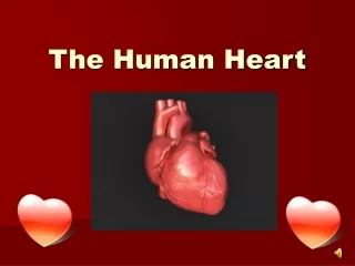
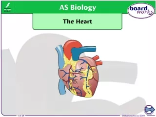
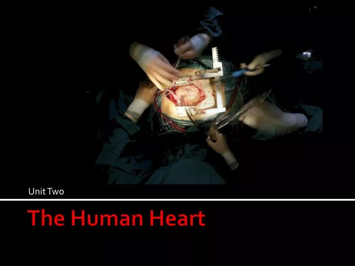
Unit Two The Human Heart
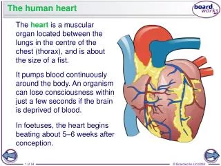
2.1.1 What is the main function? • First and foremost the human hear t is a… • PUMP! • A mechanical device using suction or pressure to raise or move liquids, compress gases, or force air into inflatable objects such as tires. • When the heart stops pumping=death • Unless the heart is restarted or intervention is used • Anna Garcia • Blood stopped flowing • Lacking the resources normally carried by the blood • including oxygen and nutrients, • Ms. Garcia’s body cells could no long survive and she died
Important pumps in our lives… • Pumps in the home: • water faucet • toilet • the washing machine • the car • the air conditioner • the refrigerator • liquid soap dispensers • spray bottles
Activity 2.1.1 • Build a simple pump using provided materials • Sketch out a few ideas • Get the OK from me to start testing • Build & Test…Trial & Error • Take GREAT notes! • Success=moving 150ml of water from one flask to the other • Explain two ways the human heart is similar to a mechanical pump. • Conclusion questions due tomorrow!
Activity 2.2.1 • Cool Heart Facts • Your heart beats about 100,000 times in one day and about 35 million times in a year. • During an average lifetime, the human heart will beat more than 2.5 billion times. • The heart pumps about 1 million barrels of blood during an average lifetime--that's enough to fill more than 3 super tankers. • Essential Question 1 • What is a pump? • Key Terms • Pump • Fluid Mechanics • Positive Displacement Pump
Activity 2.2.1: Key Terms • Histology • Inferior Vena Cava • Mitral Valve • Pericardium • Superior Vena Cava • Tissue • Tricuspid Valve • Valve • Aorta • Aortic Valve • Artery • Atrium • Cardiovascular System • Cell
Activity 2.2.1 • Three Diagrams of the heart • Exterior • Internal Ventral • Internal Dorsal Blood Circulation (Lungs/Body) • Complete first with colored pencils, markers, crayons then… • Get my OK • Add “extras” from craft room as time allows • Due Monday!
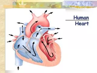
Example: Circulation • Superior Vena Cava • Inferior Vena Cava • Aorta • Right Pulmonary Veins • Left Pulmonary Veins • Right Pulmonary Arteries • Left Pulmonary Arteries • Right Lung • Left Lung • Body (head, trunk)
Let’s check our blood flow guesses? • Enters into Superior & Inferior Vena Cava • Through the Right Atrium • Tricuspid Valve • Right Ventricle • Into Pulmonary Valve • Pulmonary Artery • Thru to the Lungs • Back into Pulmonary Vein • Thru the Left Atrium • Mitral Valve (Bicuspid) • Left Ventricle • Thru the Aortic Valve • Into the Aorta • To the Circulatory System Pulmonary (lungs)& Systemic (body) Circuits http://www.youtube.com/watch?v=Rj_qD0SEGGk
We’ll come back • We’ll come back to 2.2.2. • Dissection • Microscopy • Next week • Now we’re moving on to 2.3.1
Heart Disease & Technology • Heart Disease Facts • 71,000,000 Americans have heart disease • 403 billion $$ is spent on heart disease in the U.S. • Every 34 seconds an American dies for CVD • Essential Question 1 • In what ways can technology be used to collect and analyze cardiovascular data?
Review!What Does the Cardiovascular System Do? 1. Transport _____ and ______ to all cells. 2. Remove ____ and _________ from all cells. 3. Circulate _______ for chemical regulation. 4. Help maintain body __________. (temperature, hormones, oxygen, carbon dioxide, nutrients, metabolic wastes)
Cardiac Cycle • One complete sequence of pumping/filling: • Contraction phase is called systole • Relaxation phase is called diastole • Average adult at rest completes 75 cardiac cycles per minute or 0.8 seconds per cycle • Heart Beat • Heart Attack
Electrical impulses make the Heart Beat? • SA Node (sinoatrial node) • Pacemaker • Sets timing and rhythm of heart beat • Sends electrical impulse similar to nerve impulse • Triggers cells of both atria to contract in unison • Impulse travels thorough cardiac cells to AV node (atrioventricular node)
What Makes the Heart Beat? • AV Node (atrioventricular node) • Located in wall between right atrium and right ventricle • Delays spreading the electrical impulses for 0.1 seconds to ensure the atria are completely empty • Sends impulses to specialized muscle fibers and Purkinje fibers, which conduct signal to apex of heart and induce ventricular contraction Cardiac Conduction System
Today • Activity 2.3.1 • Biomedical Science Experimental Design Protocol • Introduction to Vernier Probes
Trouble Shooting • The arrows on the two parts are pointed in the same direction. • It may be necessary to hold the receiver very close to the cylinders to initially pick-up the signal…once the signal is detected, the receiver can be moved farther away. • The receiver is within 80 cm of the hand grips. • There are no electrical devices within 25 centimeters of the receiver (including SensorDAQ, computers, cell phones, electrical lab equipment). • Have test subjects from different lab teams maintain a distance of at least 2 m from each other. • No other receivers or transmitters are near the sensor. • The contacts are clean.
Activity 2.3.2 Blood Pressure • Essential Questions • What is the relationship between blood pressure and cardiovascular function? • What factors can influence blood pressure? • Why is blood pressure important?
Activity 2.3.2 Blood Pressure • How to Write a Scientific Laboratory Report • Key Terms • Blood Pressure • Cardiology • Diastole • Diastolic Pressure • Electrocardiogram (EKG) • Hypothesis • Sinoatrial Node • Sphygmomanometer • Systole • Systolic Pressure
What Causes Blood Pressure? • Blood is a fluid • As fluids move through a pipe there is pressure exerted on the wall of the pipe= hydrostatic pressure • When blood moves through blood vessels (e.g., veins & arteries) there is pressure on the walls of the vessels= blood pressure
Blood Pressure Measurement • Pressure on arteriole wall when heart contracts= systole • Pressure on arteriole wall when heart relaxes = diastole • Blood pressure = the ratio of systolic to diastolic pressures
Blood Pressure Measurement • Traditionally measured in mm of mercury • Force that can support a column of mercury • Taken in upper arm on level with the heart • Example Measurement • 120 / 70: Systolic=120 Diastolic=70
Ever heard of MAP? • MAP= Mean Arterial Pressure • Used to measure adequacy of blood getting to vital tissues and organs • Calculated by following formula: • Systolic pressure + 2(Diastolic pressure) 3
High Blood Pressure • Clot formation • The effects of high blood pressure
Activity 2.3.2 • Part 1 (in-class today) • Part 2 (in-class today) • Conclusion questions (Due Tuesday the 25th) • Write a lab report (Due Thursday the 27th) • Lab Report Protocol • Lab Report Example • Monday is DISSECTION DAY!!!!
Activity 2.2.1 Heart Dissection • Please do not touch ANYTHING until directed • Please get into the following groups
Activity 2.2.1 Heart Dissection • Read the introduction and the opening paragraphs on page one of An Illustrated Dissection Guide to the Mammalian Heart • Use the drawings and definitions as guides to help you identify the structures of the heart. • Structures in italicsshould be located and labeled on your heart
Activity 2.2.1 External • Place the heart in your dissecting tray with the ventral side facing up. • Observe the outside of the heart. • The darker line running from the upper right diagonally to the lower left is the coronary artery. • The bottom of the heart comes to a point called the apex.
Activity 2.2.1 External • To the right and above the apex is the left ventricle. • Use your finger to push on the outside wall of the left ventricle. Notice how firm it is. • To the left and above the apex is the right ventricle. • Use your finger and push on its outside wall. Compare it to the left ventricle. Notice it compresses easier than the left ventricular wall. • Differentiate between the functions of the left and right ventricles. Use the Internet or other resources for help, if needed…(time to work on #12)
Activity 2.2.1 External • Above the ventricles is an area called the base of the heart. At each side (left and right), there are “ear like” tissue flaps called the left and right appendages, sometimes called the left and right auricles as well. • Under each appendage are the left atrium and the right atrium. • Explain the functions of the left and right atria …(time to work on #15)
Extending out of the right atrium is the superior vena cavavein. • Place a probe into it and see that it leads directly into the right atrium (this is a good strategy to be sure it is the correct structure). • Explain the function of the superior vena cava…(time to work on #17)
Activity 2.2.1 External • Next to the superior vena cava is the aorta, a large branching artery that leads to the left ventricle. • The aorta has a branch called the brachiocephalic artery. Place your finger or a probe into it and see that it leads directly into the left ventricle. • Explain the function of the aorta…(time to work on #19) • Look to the right (which is really the left), of the aorta, and see the pulmonary veins. Use a probe or your finger and see that they lead to the left atrium. • Explain the function of the pulmonary veins…(time to work on #21)
At this point it should look something like…THIS Take lots of pictures!!
Activity 2.2.1 Internal • Place the heart with the ventral side facing you. • Find the right appendage and the right atrium. • Use the scalpel to cut through the entire length of both structures. • Cut through to the cavity – not through to the other side.
Activity 2.2.1 Internal • Gently pull back the tissue exposing the inside of the cavity. • Look at the various tissues. • Use your metric ruler to measure the thickness, in millimeters, of the atrium wall (work on #28) • Observe the trabeculated (striated) lining of the appendage and the smooth lining of the atrium.
Activity 2.2.1 Internal • Cut open the superior vena cava and carefully pull back the tissue. You should see thin flaps of tissue that almost look like leaflets. This is the tricuspid valve. • Based on the name tricuspid, how many leaflets should you see? (#31) • Feel the leaflets with your finger and describe them in the space below (time to work on #32)
Activity 2.2.1 Internal • Observe the fibrous chords that are attached to the valve and help hold it in place. • These are called the chordaetendineaeand they extend to the right ventricle. • The chordaetendineae are attached to the papillary muscle, which holds the fibers to the wall of the ventricle. • Both are essential for the valve to work correctly. • Describe the function of the tricuspid valve in the space below…(work on #36)
Activity 2.2.1 Internal • Use the scalpel to make a long incision through the wall of the left ventricle. Carefully pull the wall back and observe the various tissues. • Use the metric ruler to measure the thickness of the wall of the left ventricle (in millimeters). Record the measurement: _________ (#38).
Activity 2.2.1 Internal • Compare the thickness of the wall of the left ventricle to the wall of the right ventricle. Which wall is thicker? ____________ (#39) • In the space below, describe the function of the left ventricle and explain how that relates to the difference in the wall thickness of the left and right ventricles…(work on #40)
Activity 2.2.1 Internal • Find the mitral valve(orbicuspid valve) in the left ventricle. Describe its appearance (#41) and explain its function (#42). • Check which structure is the aorta by placing your finger or a probe into it. It should lead directly to the left ventricle. • Cut open the aorta and observe the thickness of the tissue. This may also get you a better view of mitral valve. • Cut open the other major blood vessels you labeled in part one. In the space below, describe the differences you observe between the different vessels. • Based on their different functions, suggest an explanation for the differences in size and thickness of the different vessels (time to work on #43).
Activity 2.2.1 Internal • Use your scalpel to cut the heart almost in half. The cut should go through the middle but not all the way through to the other side. Leave a flap holding the organ together. • Use a probe as a pointer and starting with the superior vena cava, trace the flow of blood through the heart. In the space below, list the structures in the order the blood would meet them during its travel through the heart. Include the valves, the lungs and the extremities of your body on your list. • Reattach any labels that may have come off both hearts. Have your teacher check your dissection and your external labels.