Download
1 / 16
180 likes | 327 Views
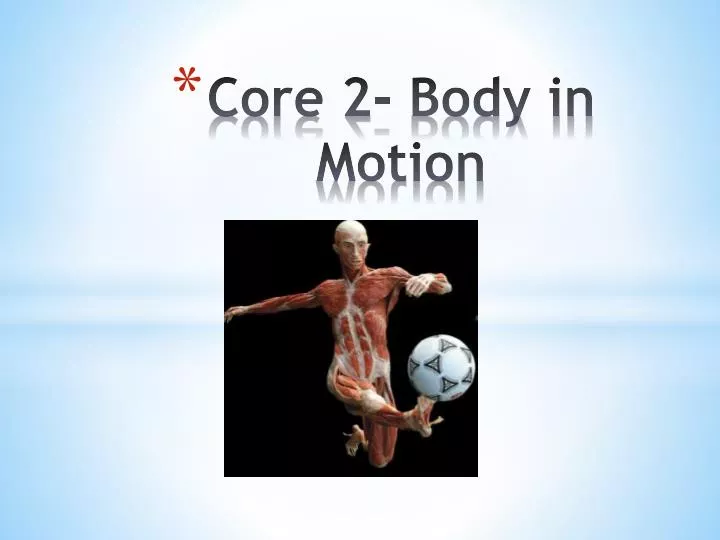
Core 2- Body in Motion. How do the musculoskeletal and cardiorespiratory systems of the body influence and respond to movement?. SKELETAL SYSTEM

E N D
How do the musculoskeletal and cardiorespiratory systems of the body influence and respond to movement? • SKELETAL SYSTEM • The human skeleton has 206 bones. They range in shape and size, a feature that allows them to perform specialised functions. The main types are long bones, short bones and flat bones. Long and short bones function as levers or to transfer forces. Flat bones usually provide protection for vital organs. • The major bones of the human body are shown in figure 4.1. Not all the bones identified in this figure are involved in movement. For example, the skull comprises many bones, but their role is to protect vital brain tissue rather than assist movement. Our focus in this chapter is on the bones that comprise and surround joints, establishing connecting structures that enable movement.
An anatomical reference system called directional terms is used to identify the location of bones. The starting point assumes that the body is in the anatomical position; that is, a reference position where the subject is standing erect, facing front on and with palms facing forward (see figure 4.2(a)). From here, we can locate a bone by saying where it is relative to another part of the body. For example, in anatomical terms, superior means ‘towards the head’. For location purposes we might say that the chest is superior to the hips or the knee is superior to the foot. Fig 4.2
Anatomical terms as they apply to locating body parts are shown in figure 4.2. • Superior — towards the head; for example, the chest is superior to the hips • Inferior — towards the feet; for example, the foot is inferior to the leg • Anterior — towards the front; for example, the breast is on the anterior chest wall (see figure 4.2(b)) • Posterior — towards the back; for example, the backbone is posterior to the heart (see figure 4.2(b)) • Medial — towards the midline of the body; for example, the big toe is on the medial side of the foot • Lateral — towards the side of the body; for example, the little toe is on the lateral side of the foot • Proximal — towards the body's mass; for example, the shoulder is proximal to the elbow • Distal — away from the body's mass; for example, the elbow is distal to the shoulder.
Bones perform five main functions in the body: 1 Support—bones provide the framework of the body; they support it and give the body shape. For example, the vertebrae support the ribs to enable us to stand. 2 Protection—bones protect vital organs within the body. For example, the pelvis surrounds the reproductive organs. 3 Movement—in conjunction with muscles, bones act as levers to allow the body to move. For example, the flexion of the knee joint allows us to kick a ball. 4 Storage of minerals—bones store minerals needed for the functioning of the body. For example, calcium is stored in bones. 5 Formation of blood cells—the formation of blood cells occurs in the cavities of certain bones.
The unique size and shape of each particular bone enable them to serve specific functions and needs in the body. Bones can be classified in the following five ways. 1 Long bones—these are long in length and elongated in shape. They consist of two ends and a shaft. They are made up of a hard shell casing (compact bone) and contain spongy bone on the inside. Examples include the femur, humerus, radius and ulna. 2 Short bones—these are cube-like and are mostly made up of spongy bone. A thin layer of compact bone provides the shape. Examples include the carpals, metacarpals, tarsals and metatarsals. 3 Flat bones— these are flat, thin bones that usually protect organs. Examples include the skull and sternum. 4 Irregular bones—these are bones that do not fall into one of the above categories; they are usually complicated in shape. Examples include the vertebrae and pelvis. 5 Sesamoid bones— these are bones found in the body where tendons pass over a joint, for example, in the foot, knee and hand. They aim to protect the tendon and increase movement. Long bones are the major bones involved in movement. They are structured as follows. Bone shaft— the long narrow part of the bone that is made of mostly marrow and compact bone Epiphysis— the head of the bone containing spongy tissue Periosteum—the thin, fibrous membrane covering the entire surface of the bone
See Figure 4.2 Classification of bones pg 93 of your textbook.
-Major bones involved in movement The skeletal system is comprised of two conjoined skeletons: the axial skeleton and the appendicular skeleton that, in concert with joints and muscles, allow movement Axial skeleton The axial skeleton provides the central structure (or long axis) of the skeletal system as shown in fi gure4.4 (see textbook). Many of the bones in the axial skeleton do not move, or move only minimally. They provide the main structure of the overall skeleton, and the core stability of the axial skeleton allows the bones of the joined appendicular skeleton (especially the long bones) to move efficiently, with both parts of the skeleton relying on the muscles that are attached to perform their appropriate function of either supporting stability or driving the movement. The axial skeleton includes the cranium, vertebral column and rib cage.
Cranium (skull) The cranium is the most complex bony structure in the body. It is formed by two sets of bones— the cranial bones and the facial bones—numbering 22 in total. The cranium bones protect the brain and organs for hearing. The facial bones form the structure of the face and cavities for the body’s senses. The skull is classified as a flat bone. Vertebral column (spine) The spine is also called the backbone of the human skeleton. It protects the spinal cord and connects the skull to the pelvis. Its 26 irregular bones (fused bones are counted as one) are classified into five sections. Cervical vertebrae—the seven vertebrae of the neck Thoracic vertebrae—the next 12 vertebrae Lumbar vertebrae—the five vertebrae supporting the lower back Sacrum—the five fused vertebrae that connect to the pelvis Coccyx—the four fused vertebrae, also known as the tailbone
Rib cage The sternum and 12 pairs of ribs make up the rib cage. All ribs attach to the posterior part of the vertebrae. Ribs provide protection around the heart and lungs. Ribs are classified as flat bones and are structured as follows. The first seven ribs are joined directly to the sternum by cartilage. The remaining five ribs are indirectly attached to the sternum; they actually join onto each other. Ribs 11 and 12 are known as ‘floating’ ribs, as they are only attached posteriorly.
The appendicular skeleton The appendicular skeleton includes all of the key long bones that are directly involved in effecting movement (see figure 4.4 of your textbook). It includes the shoulder girdle, upper limbs, pelvic girdle and lower limbs. Shoulder girdle The shoulder girdle consists of two bones: the clavicle (also known as the collarbone) and scapula (also known as the shoulder blade). The clavicle is classified as a long bone. The scapula is classified as a flat bone. These bones and surrounding muscles form the shoulder girdle. The appendicular skeleton is attached to the axial skeleton when the clavicle attaches itself to the sternum and the scapula attaches to the vertebrae. The shoulder girdle: provides attachment points for the upper limbs provides the upper limbs with flexibility and mobility not possible at any other place in the body.
Upper limbs Thirty bones comprise the upper limbs, also known as the arm, forearm and hand. The humerus, a long bone, makes up the arm segment of the upper limb. The ulna and radius make up the forearm segment of the upper arm. The ulna and humerus are responsible for the elbow joint. The radius and carpals are responsible for the hand joint; therefore, when the radius moves, the hand moves as well. Carpals are classified as short bones. Eight carpals connected by ligaments make up the wrist and five metacarpals (long bones) form the palm of the hand. The carpals, metacarpals andphalanges(fingers) make up the entire hand segment of the upper limb.
Pelvic girdle (hip) The pelvic girdle connects the lower limbs to the axial skeleton. It acts as a transfer point for weight from the upper body to the lower limbs and, as a result, plays a key role in movement. The pelvic girdle is secured to the axial skeleton by some of the strongest ligaments in the body.
Lower limbs Three segments make up the lower limb: the thigh, leg and foot. Because they carry the weight of the body when standing, the lower limb bones are thicker and stronger than bones of the upper limb. The femur, a long bone, makes up the thigh, and is the largest and strongest bone in the body. The tibia and fibula make up the leg segment of the lower limb. The tibia joins with the femur to form the knee joint, while the fibula stabilises the ankle joint. The tarsals, metatarsals and phalanges make up the foot segment of the lower limb. The foot supports and propels our body forward when we move. Seven tarsals make up the foot. The metatarsals are made up of five small long bones. In each foot, the toes are made up of 14 phalanges.
-Structure and function of joints • When two or more bones meet, they form a joint. Joints allow the body to • move and ensure that the skeleton stays together as it moves. All movements, from running, to throwing, to dancing and sitting are possible because of joints. Joints are the weakest part of the skeletal system, and are therefore susceptible to injury that may result in restricted movement. • The function of a joint is based on the amount of movement allowed. • Based on their functionality, joints can be classified as immovable, slightly • moveable and freely moveable. The structure of joints varies depending on the tissues that join the bones and whether or not there is a joint cavity. There are three ways to classify joints based on structure. • Fibrous joints—these joints are characterised by fibres joining the ends or parts of bones together. Fibrous joints use various fibres to connect bones, including short fibres (sutures), long fibres (syndesmosis) and ligaments (gomphosis). No joint cavity is present in these types of joints. These fibres make movement difficult. • Cartilaginous joints—these joints are characterised by cartilage joining the ends or parts of bones together. They contain no joint cavity and, therefore, allow only slight movement. • Synovial joints—these joints are enclosed in a capsule and covered with • cartilage and a synovial membrane to allow free movement.