Download
1 / 29
630 likes | 1.79k Views
Introduction. Incidence: 1:14,583-42,528 ER VISITS4/10 blunt laryngeal trauma expire at sceneAirwayProtectiveVoice. Anatomy and Physiology of Larynx. Airway, tracheobronchial protection,voiceHyoid, thyroid, cricoidInnervation - RLN, SLNSupraglottis - soft tissueGlottis - ca joint,cartilage,

E N D
1. Laryngeal Trauma Karen Stierman, M.D.
F.B. Quinn, M.D.,FACS
October 06, 1999
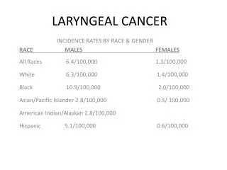
2. Introduction Incidence: 1:14,583-42,528 ER VISITS
4/10 blunt laryngeal trauma expire at scene
Airway
Protective
Voice
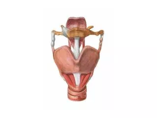
3. Anatomy and Physiology of Larynx Airway, tracheobronchial protection,voice
Hyoid, thyroid, cricoid
Innervation - RLN, SLN
Supraglottis - soft tissue
Glottis - ca joint,cartilage, neuromuscular coordination
Subglottis - cricoid, narrowest in infants
4. Anatomy and Physiology of Larynx Figure 8-60 p. 844 clin. Oriented anatomyFigure 8-60 p. 844 clin. Oriented anatomy
5. Mechanism of Injury Blunt -mva, strangulation, clothesline, cspine
Penetrating
GSW: damage related to velocity
Knife: easy to underestimate damage
6. History Hoarseness or change in voice
Dysphagia
Odynophagia
Difficulty breathing - more severe injury
Anterior neck pain
7. Physical exam Stridor -inspiratory, expiratory or both
Subcutaneous emphysema
Hemoptysis
Laryngeal tenderness,ecchymosis, edema
Loss of thyroid cartilage prominence
Associated injuries - vascular, cspine, esophageal
8. Acute Management of Laryngeal Trauma BAILEY FLOW SHEET.
AIRWAY STABILTY WILL DETERMINE WHETHER TO PROCEED WITH FLEXIBLE FIBEROPTIC EXAM OR TRACH AND PANENDOSCOPY.BAILEY FLOW SHEET.
AIRWAY STABILTY WILL DETERMINE WHETHER TO PROCEED WITH FLEXIBLE FIBEROPTIC EXAM OR TRACH AND PANENDOSCOPY.
9. Airway Management Tracheotomy under local anesthesia is preferred method for adults
CT
Fiberoptic intubation or DL with direct visualization
Pedi - inhalation anesthesia with spontaneous respirations followed by rigid endoscopic intubation
10. Radiographic Imaging C-spine
CT if airway stable and mild abnormality on flexible exam.
Good for intermediate cases with scope limited by edema
Angiography and contrast esophagrams considered
11. Medical Management Edema
Small hematoma with intact mucosa
Small glottic or supraglottic lacerations which do not involve A.C., free margin of V.C. and no exposed cartilage
Single nondisplaced stable thyroid cart. fx.
Humid. O2, airway obs., elevate HOB, H2 blockers, steroids, +/- abx.
12. Surgical Management Trach, DL, bronch, esophagoscopy
Explore within 24 hours
Lacs involving A.C. or free margin of V.C.
Large mucosal lacs, exposed cartilage
Multiple displaced cartilage fx
Avulsed or dislocated arytenoids
Vocal cord immobility
13. Laryngeal exploration and repair Bailey�s atlas p607Bailey�s atlas p607
14. Laryngeal exploration and repair Bailey�s atlas p607Bailey�s atlas p607
15. Laryngeal exploration and repair Bailey�s atlas p607Bailey�s atlas p607
16. Laryngeal exploration and repair Bailey�s atlas p607Bailey�s atlas p607
17. Goals of Laryngeal exploration Cover all cartilage to prevent granulation tissue and fibrosis
Primary closure ideal,can undermine mucosa or use advancement flaps from epiglottis or pyriforms
Palpate arytenoids and reposition if necessary
Resuspend anterior commisure, ORIF Fxs.
18. Endolaryngeal stenting Necessary for disrupted A.C., multiple displaced fractures, and/or multiple and severe mucosal lacerations
Provides support and prevents stenosis but can cause iatrogenic injury(remove after 2 weeks)
4 point fixation allows safe recovery
19. Endolaryngeal stenting Figure 68.6 BAILEYFigure 68.6 BAILEY
20. Schaefer�s classification system Looked at 139 laryngeal trauma patients over 27 years
Classified as Group I - IV and treated according to flow diagram
2/139 had poor airway on follow-up(unable to decannulate).112/115 with good voice
Time to decannulation 14-35 days,except in those patients with stents(35-100 days)
21. Schaefer�s classification system Group I - minor hematoma or lacs, no fx or airway compromise, flexible scope +/- CT, medical management
Group II -mod. edema, lacs, no exposed cart. nondisplaced fx. varying airway,trach +/- CT
Group III - Massive edema, disrupted mucosa, displaced fx, cord immobility, varying airway, trach and endoscopy
Group IV multiple unstable fx, a.c. trauma, required a stent
22. Special considerations LT separation - usually immediate death,if not: trach then suture cricoid to 2nd tracheal ring. Assoc. with BRLN injury and stenosis
RLN injury - direct repair if possible but poor chance for functional return
Pedi - Proportionally smaller airway tolerated less edema however pedi larynx more flexible so more soft tissue injury
23. Complications Granulation tissue - most common, prevention key, can lead to fibrosis and stenosis of larynx or trachea, tx is site specific and includes laser excision, laryngofissure and cricoid split
Immobile vocal fold - cricoarytenoid joint or RLN injury. If arytenoid mobile, may observe for return of nerve function
24. Conclusions Key to recognition is high index of suspicion
Assess airway first and base management on flow diagram
Don�t forget about associated vascular or esophageal injuries
25. Case presentation 92 yom s/p MVA presented to ER c/o pain in neck and hoarseness
26. Physical exam Anterior neck contusion and hematoma
Pain with palpation of larynx
27. Fiberoptic exam Unable to see mucosa or cartilage disruption but the larynx seems somewhat abnormal in appearance
28. CT scan Fx of the thyroid cartilage posterior and laterally with some displacement, fx of midline thryoid cartilage
29. Management Trach/DL/esophagoscopy, laryngeal thyrotomy with repair of unstable fx and mucosal lacerations.