Download
1 / 18
180 likes | 189 Views
Pb(II) and Hg(II) binding to de novo designed proteins studied by 204m Pb- and 199m Hg-perturbed angular correlation of γ-rays (PAC) spectroscopy: Clues to heavy metal toxicity L. Hemmingsen, Th. Agne, S.K. Das, S.-B. Ruy, V. L. Pecoraro , J.G. Correia. Outline

E N D
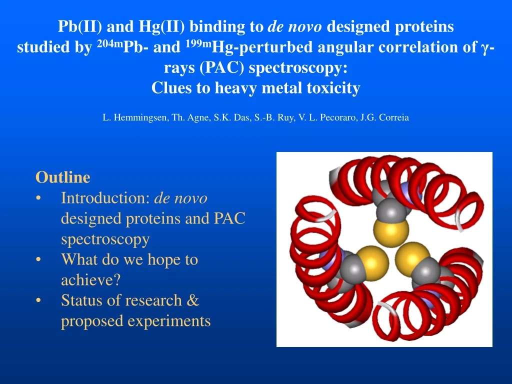
Pb(II) and Hg(II) binding to de novo designed proteins studied by 204mPb- and 199mHg-perturbed angular correlation of γ-rays (PAC) spectroscopy: Clues to heavy metal toxicity L. Hemmingsen, Th. Agne, S.K. Das, S.-B. Ruy, V. L. Pecoraro, J.G. Correia • Outline • Introduction: de novo designed proteins and PAC spectroscopy • What do we hope to achieve? • Status of research & • proposed experiments
De novo designed proteins: Heptad repeats 1 10 20 30 g a b c d e f g a b c d e f g a b c d e f g a b c d e f g a G-LKALEEK-LKALEEK-LKALEEK-LKALEEK-G heptad #1 heptad #2 heptad #3 heptad #4 • Inspired by naturally occurring • coiled coils • Hydrophobic core of leucines
De novo designed proteins: Structure determined by X-ray diffraction • Tripple-stranded α-helical bundle • Hydrophobic core of leucine side • chains – as expected • Initially used for studies of protein • folding Lovejoy et al. Science 1993, 259:1288
De novo designed metalloproteins: The L16C TRI-peptide series L16CG-LKALEEK-LKALEEK-CKALEEK-LKALEEK-G UV absorption spectroscopy of Cd(II) binding Matzapetakis et al. J. Am. Chem. Soc., 2002, 124:8024
De novo designed proteins offer a structural framework for... • Creation of natural metal ion binding sites, to test essential • features necessary for: • Electron transfer • Metal ion regulated processes • Metalloprotein folding, structure, dynamics, and • catalysis • Creation of metal ion binding sites with properties • unprecedented in nature • The aim of this work is to create and characterize • heavy metal ion binding sites mimicking those found in • nature – hoping to capture the molecular basis of heavy • metal toxicity
Perturbed angular correlation of γ-rays: The influence of extra-nuclear fields The nuclear quadrupole interaction is proportional to: Thus, ω1, ω2, and ω3 represent a fingerprint of the local charge- distribution eQ 111Cd Hamilton Phys. Rev., 1940, 58:122, Brady and Deutsch Phys. Rev.1947, 72:870, Goertzel Phys. Rev., 1946, 70:897, Aeppli et al. Phys. Rev., 1951, 82, 550, Leipert et al. Nature, 1968, 220:907
PAC in a nutshell W180W90 Calculate spectroscopic data based on structure Compare Hemmingsen et al. Chem. Rev., 2004, 104:4027
L16C Combination of PAC, NMR, and QM calculations: An exchanging water molecule 113Cd NMR Matzapetakis et al. J. Am. Chem. Soc., 2002, 124:8024
L16C Combination of PAC, NMR, and QM calculations: An exchanging water molecule 113Cd NMR PAC spectroscopy provides unique information on structure and dynamics
What do we hope to achieve? • Fundamental knowledge of Pb(II) and Hg(II) interaction with proteins mimicking naturally occuring binding sites: • Structure and dynamics at de novo designed metal ion binding sites • Selectivity and affinity for these heavy metals • Clues to the molecular basis of toxicity of these heavy metals • Biotechnological long term goal: Design of high affinity • high selectivity heavy metal chelators
De novo designed proteins: Pb(II) binding to a and d sites • UV-Vis-, CD- and EXAFS spectroscopic studies indicate that Pb(II) binds preferentially to d sites contrary to Cd(II) and Hg(II) • Unfortunately, assignment of the spectroscopic signals to a and d sites has not been possible
De novo designed proteins: Pb(II) binding to a and d sites 1. Using L16C (a site) and L12C (d site) we will assign 204mPb- PAC signals to the two sites, and characterize the two sites in terms of structure 2. In proteins with two binding sites (separated by one leucine layer) we will study selectivity for Pb(II) versus Cd(II) and Hg(II)
3 – 4.5 eq CSL9C 0 – 2 eq CSL9C De novo designed proteins: Hg(II) binding to CSL9C UV absorption (and 199Hg NMR) spectra display a distinct change from addition of 0-2 eq. CSL9C to addition of 2-3 eq. CSL9C to a Hg(II) solution – does this reflect formation of a two-coordinated Hg(CS-L9C)2 complex and a three-coordinated Hg(CSL9C)3-, respectively?
3 – 4.5 eq CSL9C 0 – 2 eq CSL9C De novo designed proteins: Hg(II) binding to CSL9C PAC spectroscopy should allow for discrimination of two- and three- coordinated structures, with considerably higher frequencies for the linear complex sub-stoichiometric studies is a particular advantage of PAC- spectroscopy
3 – 4.5 eq CSL9C 0 – 2 eq CSL9C De novo designed proteins: Hg(II) binding to CSL9C Is the (potential) three-coordinated structure trigonal planar or T- shaped? PAC spectroscopy is ideally suited for discrimination of these two structures, with an asymmetry parameter of 0 in the prior and 1 in the latter case
De novo designed proteins: Hg(II) binding For all the peptides the following equilibrium appears to exist: Hg(peptide)3 (= HgS2SH) ↔ Hg(peptide)3- (= HgS3) + H+ Does the protonated cysteine coordinate to Hg(II)? PAC spectroscopy is expected to solve this problem as the electric field gradient will depend strongly on the charge state of the SH group
De novo designed proteins: Hg(II) binding Proteins have been designed with two metal ion binding sites next to each other (6 cysteines in total) presenting variety of possible coordination geometries PAC spectroscopy should be well suited for discrimination of 2-, 3-, and 4-coordinated Hg(II) ions with thiolate ligands
Summary • De novo design of proteins is a powerful tool in the study of fundamental heavy metal – protein interactions • PAC spectroscopy provides unique information on structure and dynamics of metal ion binding sites • We hope to uncover fundamental chemistry of Pb(II) and Hg(II) interaction with proteins: • Structure and dynamics at de novo designed metal ion binding sites • Selectivity and affinity for these heavy metals • Clues to the molecular basis of toxicity of these heavy metals • Biotechnology: Design of heavy metal chelators