Download
1 / 28
290 likes | 609 Views
First Trimester Ultrasound. Questions. What Measurement is the most accurate for ultrasound dating? In what order do fetal structures appear? What are the 7 items to document in 1 st trimester ultrasounds?. Objectives. Review the indications for first trimester ultrasound

E N D
Questions • What Measurement is the most accurate for ultrasound dating? • In what order do fetal structures appear? • What are the 7 items to document in 1st trimester ultrasounds?
Objectives • Review the indications for first trimester ultrasound • Discuss utilization of ultrasound and laboratory data in the evaluation of first trimester bleeding • Review measurements and how they apply to dating criteria • Discuss how to document a first trimester ultrasound inthe medical record
Indications • Dating of pregnancy • Size vs dates discrepancy; multiple gestation determination *(1st Trimester)* • Vaginal bleeding • Abdominal or pelvic pain: rule out ectopic pregnancy/ torsion/ heterotopic pregnancy/ ovarian cyst ***Not credentialed to do*** • To confirm viability
Pregnancy Dating with 1st trimester ultrasound • The only utility for “routine” ultrasound as determined by the RADIUS study • Early dating is the most accurate (+/- 5-7d or 8%) • Better defines timing for later testing and interventions • Triple/Quad test • Tocolysis/Steroids • Reduces the incidence of induction for postdates
Measurements • Mean Sac Diameter • Should be measured in 3 dimensions • May be all that is visible at the discriminatory zone; IUP best confirmed with some fetal element, such as a yolk sac • Embryonic Crown-Rump Length (CRL) • Measurement of a CRL with fetal cardiac activity is the best measurement for dating purposes
Typical Measurements • There are tables for determining gestational age based on: • Gestational Sac Measurement • Crown-Rump Length • All of the U/S machines at NHP contain software which perform these calculations. The measurements will trigger the gestational age determination.
Early Pregnancy Failure • Failure of appropriate interval growth by u/s of embryo • Fetal pole/yolk sac should be seen by the time the MSD is 20 mm (not as accurate as FCA though) • Fetal Cardiac Activity should be seen by the time the CRL is 4mm (5mm per AIUM) • If not, may repeat the u/s in one week
Rule out ectopic • Classic triad—amenorrhea, vaginal bleeding, pain • Must have a high index of suspicion • Even more so in the face of risk factors • Three primary tools for evaluation • Physical exam • Quantitative β HCG • Ultrasound
Lab and Ultrasound • Discriminatory Zone—the quant β-hCG level at which one would expect to be able to identify an intrauterine pregnancy • For vaginal sonography—1200-1500 (1000-2000 per ACOG) • For abdominal sonography—3000-4000 • If the quant β-hCG is at or above the discriminatory zone, AND no IUP can be identified, the pregnancy may be ectopic
Rapidly Rising Quant β HCG • Identifying multifetal gestation • Identifying gestational trophoblastic disease • Don’t forget lab error (i.e. normal pregnancy in your differential)
Other applications • Evaluation of gynecologic structures • Uterus—position, fibroids • Adnexae—masses, corpus luteum • Early screen for chromosomal anomalies • Nuchal translucency measurements
Documentation • Whether obtained abdominally or vaginally, the following information should be obtained and documented: • Presence or absence of IU gestational sac and identification of an embryo if possible • Fetal number • Presence or absence of fetal cardiac activity • Crown-rump length • Evaluation of uterus and adnexal structures and presence of free fluid
Final Pearls • Do not include the yolk sac with the CRL • Practice, practice, practice • Abdominal and Vaginal • If you are not sure it is an IUP, get help
What Measurement is the most accurate for ultrasound dating? • Crown Rump Length – Up to the 12th week of life.
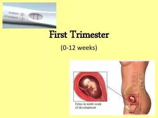
In what order do fetal structures appear? • Gestational sac – 4 to 5 weeks • Yolk sac – 5 to 6 weeks • Fetal pole - 6 to 7 weeks • Cardiac Activity - 6 to 7 weeks.
What are the “lucky #7” components of a first trimester ultrasound again?
Components of documentation of a first trimester ultrasound • #1: IUP or no IUP • #2: how many fetuses • #3: FCA or no FCA • #4: CRL • #5: uterine masses • #6: adnexal masses • #7: free fluid 27
The end 28