Download

1 / 12
130 likes | 378 Views
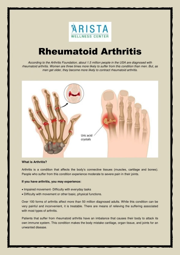
Tests for Rheumatoid Arthritis. Chua, Kathleen. Laboratory Findings. Rheumatoid factors Antibodies to Cyclic Citrullinated Peptide (Anti-CCP) CBC with differential count Erythrocyte sedimentation rate Synovial Fluid Analysis. What are Rheumatoid Factors ? .

E N D
Tests for Rheumatoid Arthritis Chua, Kathleen
Laboratory Findings • Rheumatoid factors • Antibodies to Cyclic Citrullinated Peptide (Anti-CCP) • CBC with differential count • Erythrocyte sedimentation rate • Synovial Fluid Analysis
What are Rheumatoid Factors? • Autoantibodies reactive with the Fc portion of IgG • Its presence is NOTspecific for RA • Frequency increases withage • 10-20% of individuals >65 y/o test positive despite not having RA
Other conditions associated with the presence of rheumatoid factors: • Systemic Lupus Erythematosus • Sjögren’s Syndrome • Chronic liver disease • Sarcoidosis • Interstitial pulmonary fibrosis • Infectious mononucleosis • Hepatitis B • Tuberculosis • Leprosy • Syphillis • Subacute bacterial endocarditis • Visceral leishmaniasis • Schistosomiasis • Malaria • Healthy individuals • > 65 years old • Post-vaccination • Post-transfusion • Relatives of patients with RA
Rheumatoid factor is used only to evaluate.It is NOT used as a screening procedure • Predictive value of the presence of rheumatoid factor in determining a diagnosis of RA is poor • < 1/3 of patients with (+) rheumatoid factor will be found to have RA • Evaluate severity and progression • ↑ titer = ↑ severity
Anti-Cyclic Citrullinated Peptide(Anti-CCP) • Most commonly found in patients with: • (+) rheumatoid factor • Aggressive disease bone errosions • RA-associated HLA-ß1 allele • Smokers • Early detection of RA • Sensitivity: anti-CCP = rheumatoid factor • Specificity:anti-CCP > rheumatoid factor • Confirm diagnosis, establish prognosis • Disadvantage vs. rheumatoid factor: not useful in predicting the future development of RA • Can’t evaluate severity and progression
Hematologic Findings • Complete Blood Count w/ Platelet Count and Differentials • Normochromic, normocytic anemia • ineffective erythropoiesis • Thrombocytosis • Usuallynormal WBC count but… • May have mild leukocytosis • Felty’s syndrome – leukopenia • Severe systemic disease – eosinophilia • ↑iron stores in bone marrow • ↑ erythrocyte sedimentation rate
Other serologic findings • Acute phase reactants • Ceruloplasmin • C-reactive protein Correlate with disease activity and likelihood of progressive joint damage
Synovial fluid analysis • Confirms presence of inflammatory arthritis • Non-specific • Characteristics of synovial fluid • Physical • Turbid • Reduced viscosity • Clinical chemistry • Increased protein content • Normal / slightly decreased glucose concentration • Cell count • WBC varies from 5 to 50,000/uL; PMNs predominate • >2000/uL with > 75% PMNs = inflammatory arthritis • Non-diagnostic of RA • C3 & C4 markedly diminished
Radiographic Evaluation • None of the radiographic findings is diagnostic of RA • Early in the disease – usually NOT helpful; will only reveal the “obvious”, not help significantly in management • As the disease progress – abnormal findings more pronounced
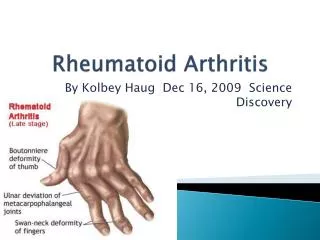
Characteristic patterns of abnormalities seen in radiographs • Symmetric involvement • Juxtaarticularosteopenia • Loss of articular cartilage • Radiography would determine the extent of cartilage destruction • Bone erosion X-rays of the hands in Rheumatoid arthritis. Demonstrates periarticularporosis, joint space narrowing of the proximal interphalyngeal joints, and erosions. Note erosion of the ulnarstyloid, and narrowing of the wrists.
Other imaging procedures • 99mTc bisphosphonate bone scanning • MRI Technetium-99 bone scan in a patient complaining of stiffness and painful joints but a NORMAL examination, showing uptake of technetium in sub clinical inflammation of joints. Note symmetrical, polyarticular uptake pattern (hands, feet and knees demonstrated)- typical of Rheumatoid arthritis.