Download
1 / 92
940 likes | 1.04k Views
Anemia. Goals. Definition of anemia . Epidemiology. Classification. Review the terminology used to discuss anemia. Evaluation: . • Address clues in the history and exam that can direct the evaluation. • Describe one approach to using lab tests to make the diagnosis.

E N D
Goals • Definition of anemia . • Epidemiology. • Classification. • Review the terminology used to discuss anemia. • Evaluation:. • Address clues in the history and exam that can direct the evaluation. • Describe one approach to using lab tests to make the diagnosis. • Review some anemia& aspects of treatment of common anemia.
Definitions A.Anemia refers to a reduction in the number or volume of RBCs to less than a normal level. May occur due to: • Acute/chronic blood loss • Decreased production • Breakdown of blood (hemolysis)
B. Strictly defined by decreased Hgb relating to a value 2SD below a mean .
2 . Specific lab values must be interpreted in context of : • patient and illness; ie, are Hgb levels of 14.0 in a male smoker, or of 12.5 in a severely volume contracted woman, “normal?”
C. The clinical context and condition of the patient determine how urgently Dx and Rx must occur.
EPIDEMIOLOGY • Anemia affects more than 2 billion people worldwide – one-third of the world's population. • Half of these are iron deficiency anemia. • Anemia is common in menstruating women,infant, children and elderly patients. • In almost all developing countries, at least 1/3 of the Women and children are anemic. • Prevalence among pregnant women and children under 2 years of age is more than 50 %. • Anemia contributes to 20% of all maternal deaths.
Indices • 1. Hgb – most reliable value • 2. Hct – reliable if spun; if automated it is an approximation. • 3. MCV (mean corpuscular volume) – is a useful index for distinguishing anemias (if homogenous RBC population). • • Normal MCV 82-97 fL • • Macrocytosis > 97 fL • • Microcytosis < 82 fL
Mentzer index = MCV/RBC Ratio <13: Thalassemia Ratio >13: Iron Deficiency Anemia, Hemoglobinopathy. • Mean corpuscular hemoglobin (MCH) - Average quantity of Hb per red cell MCH (pg) = (Hb x 10) ÷ RBC
4. MCHC (mean corpuscular Hgb concentration) – derived index Grams of hemoglobin per 100 cc packed cells. MCHC = (Hb ÷ PCV) x 100 • 5. RDW (red blood cell distribution width) – calculated index (nml 11.5-14.5%); if elevated, indicates variability of RBC (anisocytosis) – unreliable value in diagnosis. It measures RBC size variation (Anisocytosis) Standard deviation of RBC volume x 100 MCV • It is very helpful to distinguish between different types of microcytic anemia.
Reticulocyte Count: Decreased: 1- Hypoproliferative 2- ineffective erythropoiesis Increased : 1 -Iron deficiency anemia 2 Lead poisoning 3 Condition with high reticulocyte count (thalassemia minor?) 4 Erythropoieticporphyria 5 Inflammation
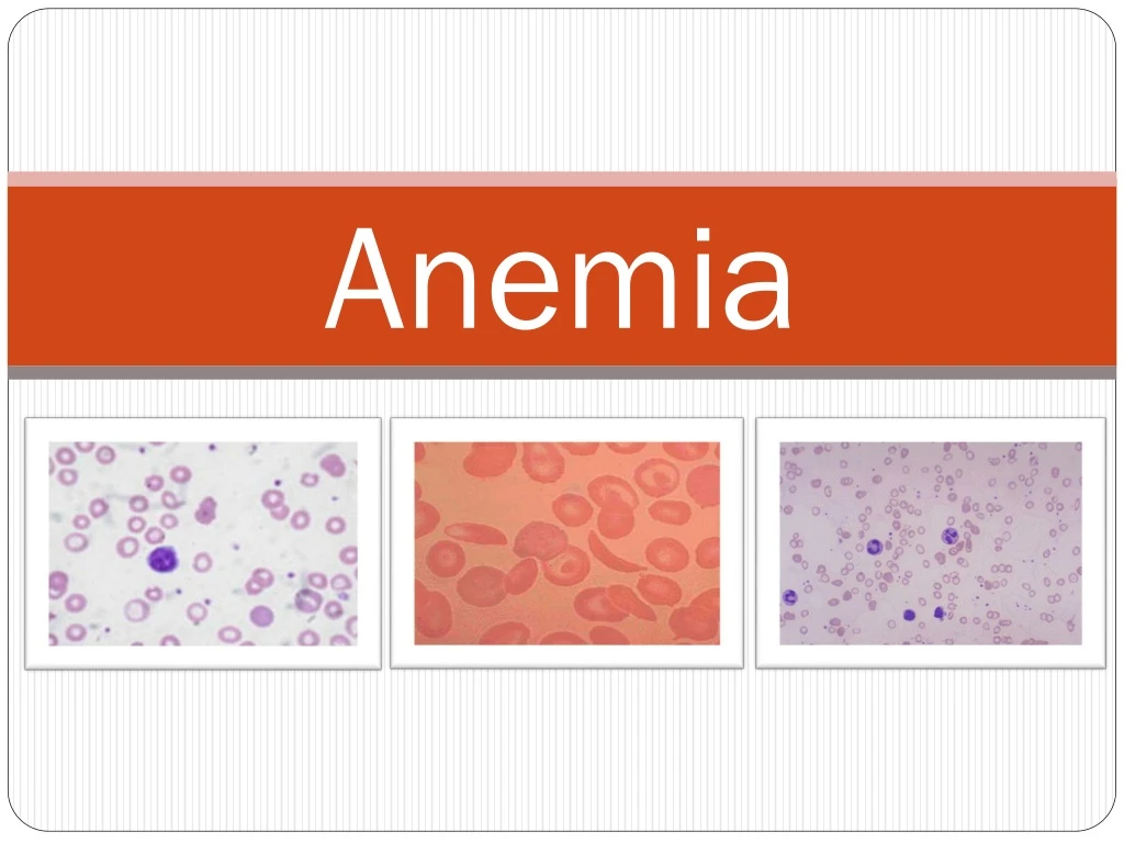
E. Remember the value and often the need, to actually examine the peripheral smear • Spherocytes/ovalocytes • Sickle cells • Schistocytes – traumatic hemolysis (prosthetic valve, DIC, TTP, hemolytic-uremic syndrome) • “Teardrop” cells – bone marrow disease (fibrosis, tumor) • Oval macrocytes, hypersegmentedpolys – megaloblastic anemia • Immature (blast) cells – leukemia
classification Morphologic • MCV,MCHC,RDW Functional • Decrease production • Increase destruction Underlying mechanism • Impaired production(proliferation ,differentiation ,maturation ) • Increased destruction-hemolytic. • Blood loss.
Anemia Macrocytic anemia (MCV>100) Microcytic anemia (MCV<80) Normocytic anemia (MCV 80–100) With megaloblastic bone marrow Without megaloblastic bone marrow High reticulocyte count Low reticulocyte count
Functional classification I. inadequate production of RBCs : A. deficiency of essential factors B. toxic factors C. endocrine causes (thyroid ,pituitary) D. erythropoiten production (renal, starvation) E. invasion of bone marrow f. failure of stem cells II. Increased RBC loss or destruction(hemolytic anemia) III. Blood loss anemia
A.History 1. Hx of chronic anemia or family Hx of anemia – may suggest inherited anemia. • Spherocytosis, ovalocytosis • Hemoglobinopathy 2. Medical Hx – many chronic illnesses can be associated with anemia. • Chronic infection • Diabetes, hypothyroid, renal, hypoadrenal, collagen vascular diseases are common causes. • Malignancy
3. Social Hx • Nutritional: strict vegan (B12); few fruits/veggies (folate) • Alcohol use (folate, marrow suppression, liver disease) . 4. Surgical Hx: partial or total gastrectomy.
5. Medications can cause • Bone marrow depression • Hemolysis (G-6-PD deficiency) 6. Review of systems • Pregnancy • Menses • Symptoms suggesting undiagnosed medical problem
B. Symptoms attributable to anemia alone 1. Usually not present until Hgb level less than 7-8 g/dl. 2. No correlation between level and signs/symptoms.
C. Physical findings 1. Pallor of oral mucosa/conjunctiva 2. Palmar crease pallor suggests Hgb < 7 g/dl.
3. Volume status: orthostasis, baseline tachycardia, widened • pulse pressure, flow murmurs, flat neck veins, decreased urine output, decreased turgor. 4. Skin: jaundice (hemolysis), petechiae/ecchymoses (bleeding disorders), lymphadenopathy(malignancy/infection)
5. Oral • Glossitis, macroglossia (pernicious anemia) • Angular cheilitis (Fe deficiency) Angular Stomatitis Glossitis
Angular Stomatitis Koilonychia
6.Neuro: paresthesias, dementia, ataxia, decreased proprioception/vibration (pernicious anemia). 7. Heme positive stool (GI loss). 8. Splenomegaly (hemolysis, sequestration, malignancy).
Symptoms • The symptoms can be related to the anemia itself, or the underlying cause: • fatigue. • Dizziness. • headache. • poor concentration • Parasthesia in fingers and toes. • Irritability • dyspnea. • increasing cardiac output: palpitation , intermittent claudication and symptoms of heart failure .
D. Labs 1. Approach varies greatly depending upon reference, experience, circumstances, etc; regardless of approach, have a rationale for it. • Recognize the difference between patient in office vs in hospital (usually acutely ill). • Try not to “shotgun” (even though we all do it!)
2. Approach a. CBC, peripheral smear b. Check retic count 1) Must calculate the corrected absolute retic count • Abs retic count = retic count x RBC • Abs retic count < 100,000 suggests defect inRBC production • Abs retic count > 100,000 suggests acute bleeding or hemolysis
2) Corrected Abs retic count = Abs retic count/retic maturity time • Hct 45% Mat time 1 day • Hct 25%, 2.0 days • Hct 15%, 2.5 days
Anemia Macrocytic anemia (MCV>100) Microcytic anemia (MCV<80) Normocytic anemia (MCV 80–100) With megaloblastic bone marrow Without megaloblastic bone marrow High reticulocyte count Low reticulocyte count
3). Specific notes a. Serum Fe: negative phase reactant – it decreases with any stress (fever, etc). b. TIBC: only elevated in Fe def; however, it is also a negative acute phase reactant. c. % Sat: decreases in both Fe def and ACD
d. Ferritin: proportional to body’s iron stores generally both increase with age. • Less than 16-35 ng/dl suggests depleted stores (if older than 65, less than 45 ng/dl). • Even though it is a positive acute phase reactant, must have Fe to elevate. • Can have ferritin of 50-60 ng/mL and still have Fe deficiency. e. Bone marrow iron stores: (gold standard).
MicrocyticAnemias DIFFERENTIAL DX: • Iron Deficiency Anemia • Thalassemias • Sideroblastic Anemia • Lead poisoning • Anemia of chronic disease (usually normocytic) MCV < 80 Check also MCH, MCHC, RDW Next Lab testsIron studies Serum Iron TIBC/transferrin Serum Ferritin, % sat
MicrocyticAnemia (MCV < 82 fL) • 1. Check ferritin level • a. Low value (generally < 30 ng/mL) suggests/confirms iron def. • b. Normal or high value – check serum iron. • 1) Low Fe – anemia of chronic disease (ACD) • 2) Normal or increased – check serum lead level. • High – lead toxicity • Normal – do Hgb electrophoresis: thalassemia
Iron Deficiency Anemia Most common cause of anemia worldwide 1. Prevalence:10-25% young women, 1% men, up to 10% elderly 2. Why worry? a. Treatable b. Clue to underlying diseases • 10-20% pts w/Fe def anemia have CA. • Up to 50% have GERD/PUD 3. Diet 10-15 mg/day – 10% absorbed
4. Daily loss 1 mg/d, plus 1 mg/d menstruation 5. FeSO4 20% elemental iron: 180 mg = 36 mg elemental Fe 6. Replace Fe at 6 mg/kg/day up to 200 mg/d elemental Fe 7. Consider Feosol elixir to minimize GI side effects common with FeSO4 tablets.
Iron deficiency anemia • Iron deficiency anemia is the most common type of anemia
8. Vitamin C increases absorption of non-heme Fe; literature implies little clinical significance, but antioxidant Rx is becoming a hot topic. 9. Retic count up by 2 weeks 10. Anemia corrected by 6 weeks 11. 4-6 months to correct depleted Fe stores of 500 mg 12. In questionable cases, especially of distinguishing Fe-def vs ACD in an elderly ill patient, consider empiric trial of Fe replacement. Be sure to follow retic and Hgb; if no change, stop Fe
PREVALENCE OF ID • Region0-4yr 5-12yrWomen • South Asia 56% 50% 58% • Africa 56% 49% 44% • Latin Am 26% 26% 17% • Gulf Arabs 40% 36% 38% • Developed 12% 7% 11% World43% 37% 35%
Causes of iron-deficiency anemia 1. Increased need • Pregnancy • Normal growth 2. Decreased intake or absorption • Childhood • Gastric surgery, achlorhydria • Celiac sprue 3. Increased blood loss • GERD, PUD, gastritis • IBD • Malignancy • Menstruation
Clinical Signs to be looked for • Skin / mucosal pallor, • Skin dryness, palmar creases • Bald tongue, Glossitis • Mouth ulcers, Rectal exam • Jaundice, Purpura • Lymph adenopathy • Hepato-splenomegaly • Breathlessness • Tachycardia, CHF • Bleeding, Occult Blood