Download
1 / 15
170 likes | 554 Views
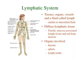
Lymphatic System. Chapter 20. An Overview. One way system flowing towards heart Functions Return fluid and proteins to venous blood House phagocytic cells and lymphocytes Carry absorbed fats from intestines to blood Components Lymphatic vessels Lymph Lymph nodes. Lymphatic Vessels.

E N D
Lymphatic System Chapter 20
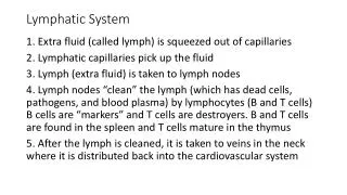
An Overview • One way system flowing towards heart • Functions • Return fluid and proteins to venous blood • House phagocytic cells and lymphocytes • Carry absorbed fats from intestines to blood • Components • Lymphatic vessels • Lymph • Lymph nodes
Lymphatic Vessels • Capillaries • Endothelial cells form minivalves • Inflammation increases permeability • Lacteals carry fat from intestines as chyle • Collecting Vessels • Similar to veins • Varies between individuals • Trunks • Lumbar • Bronchomediastinal • Subclavian • Jugular • Intestinal • Ducts • Right lymphatic • Thoracic • Cisterna chyli • Dump to venous blood
Lymph Transport • Low pressure system w/o a pump • Similar return as veins • Arterial pulsations • Tunica media smooth muscle contraction • Balances with blood fluid loss • Hydrostatic and colloid pressures (Chpt. 19) • ~ 3L every 24 hours • Rate increases w/activity
Lymphocytes • Primary fighters of immune response • Targets are antigens • T-cells – direct attack • Attack and destroy antigens • B-cells – indirect attack • Produce antibodies from plasma cells to ‘flag’ antigens
Other Lymphoid Cells • Macrophages • Phagocytic themselves • Activate T-cells • Dendritic cells • Capture and move antigens to lymph nodes • Activate T-cells too • Reticular cells • Fibroblast-like cells that form supportive network
Lymphoid Tissue • Proliferation & surveillance sites • Primarily reticular CT (except thymus) • Diffuse lymphatic tissue • Sparse scatterings in all lymph organs, • Concentrated in lamina propria of mucus membranes • Lymphoid follicles (nodules) • Spherically packed tissue w/o capsule • Larger organs and few isolated patches • Germinal centers where B cells proliferate • Enlarge w/ increased B cell division
Lymph Nodes • Main lymphatic organs • Located along lymph vessel path • Concentrated near large collecting vessel junctions • Inguinal region • Axillary region • Cervical region • Functions • Filtration • Macrophages prevent foreign molecule entrance to blood • Immune system activation • Monitor for antigens to fight
Lymph Node Structure • Dense fibrous capsularoutside • Difference b/w node and nodule • Invaginatesforming trabeculae • Regions • Medulla • Macrophages, T cells, B cells, and plasma cells • Lymph sinuses: capillaries where macrophages ‘hunt’ • Leaking antigens activate lymphocytes in tissue • Cortex • Dense nodules w/germinal centers • Transient T cells
Lymphatic Circulation • Enters node in afferent lymphatic vessels • Large subscapular sinus to smaller, cortical sinuses • Enter medulla • Exit at hilum via efferent lymphatic vessels • Fewer slows flow • Allows lymphocytes & macrophages to work
Spleen • Largest lymphatic organ • Functions • Lymphocyte proliferation and surveillance • Stores products of RBC breakdown and platelets • Cleanse blood • Remove aged/damaged blood, debris, and foreign matter • Fetal erythrocyte production (ceases after birth) • Distinct areas • White pulp w/lymphocytes act in immune functions • Red pulp w/worn out erythrocytes and pathogens
Thymus • Bilobed organ at base of neck • More pronounced when young • Corresponds w/importance of immune function • T lymphocyte maturation only • Lacks B cells • Doesn’t directly fight antigens • Thymocytes secrete thymosin and thymopoietin to signal T cell maturation
Tonsils • Lymphatic tissue ring around pharynx • Palatine: largest and most likely infected • Lingual • Pharyngeal (adenoids) • Tubal • Follicles w/germinal centers • Gather and remove pathogens from pharynx • Crypts are deep invaginations to trap and destroy • Tonsil stones • Produces ‘memory’ immune cells for future attacks
Mucosa-Associated Lymphatic Tissue (MALT) • Collections of lymphatic tissue to protect external environment openings • Peyer’s patches • In walls of small intestine • Destroy bacteria before it leaves intestines • Generate ‘memory’ lymphocytes • Appendix • Junction of small and large intestine • Similar function as Peyer’s patches • Lymphoidnodules • In walls of bronchi
Homeostatic Imbalances • Tonsillitis: inflammation of tonsils • Lymphangitis: vasa vasorum of lymph vessels congested w/blood • Lymphedema: blockage prevents return to blood • Buboes: inflamed lymph nodes • Splenectomy: removal of a ruptured spleen • Appendectomy: removal of appendix • Elephantiasis: lymph vessels clogged by worms causing increased swelling • Hodgkin’sdisease: malignant B-cells • Non-Hodgkin’slymphoma: any lymphoma, but Hodgkin’s