Download
1 / 47

E N D
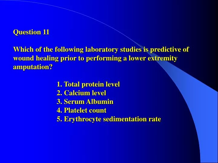
Question 11Which of the following laboratory studies is predictive of wound healing prior to performing a lower extremity amputation? 1. Total protein level 2. Calcium level 3. Serum Albumin 4. Platelet count 5. Erythrocyte sedimentation rate
Answer: 3 L. KroonenSerum albumin < 3.0 g/dl and total lymphocyte count of < 1500 both are associated with poor wound healing. Above these levels, an 82% healing rate is quoted.Another thing to consider is the vascular status of the foot. Toe pressures of greater than 45mmHg and transcutaneous PO2 greater than 30 are both correlated with good wound healing. PO2 less than 20 makes wound healing unlikely.Hemoglobin greater than 10g/dl will provide sufficient O2 to the wound.Ischemic index of 0.5 or greater will support wound healing. This is the ratio of the Doppler pressure at the level being tested to the brachial systolic pressure.Richardson, EG. Amputations about Foot. In Campbell’s Operative Orthopedics, Chapter 10, pg 555.Gottschalk, FA. Rehabilitation: Gait, Amputations, Prosthetics, Orthotics. In Miller (ed.) Review of Orthopaedics. pp505-6.
Question 18 - When harvesting the flexor digitorum longus distally in the foot, which of the following tendons crosses immediately below it? 1. Flexor hallucis longus 2. Flexor hallucis brevis 3. Adductor hallucis 4. Abductor hallucis 5. Quadratus plantae
Answer: 1M Harpe Anatomy question, although poorly worded . . . Rather than stating “immediately below it” the question should have been worded “deep to it” . . . In anatomic position reference, the Flexor hallucis longus tendon passes immediately deep to the Flexor digitorum longus . . . None of the other choices would be appropriate.
Question 28 Which of the following is considered the most common cause of recurrent ankle sprains in dancers? 1. Poor proprioception 2. Hypermobility of the subtalar joint 3. Lateral ligament laxity 4. Peroneal tendon weakness 5. Posterior tibial tendon dysfunction
Clarke Answer: 4 Peroneal injury including tendinopathy and partial tears of both the peroneus longus and brevis tendons has been linked to persistent lateral ankle pain and chronic lateral ankle instability. Di Giovanni et al report on 61 patients who underwent a primary ankle ligament reconstruction for chronic instability. Associated injuries found at surgery included 77% of patients with peroneal tenosynovitis, 54% with attenuated peroneal retinaculum, and 25% with peroneus brevis tear. Based on their experiences, both Di Giovanni et al and Alanen et al suggest that peroneal tendon injuries may be an often overlooked cause of persistent lateral ankle pain and chronic ankle instability in settings of overuse and after acute trauma. Hamilton et al. Foot and Ankle injuries in dancers. Surgery of the Foot and Ankle, ed 7. St Louis, MO, Mosby, 1999. 1225-1256. Hamilton. Foot and Ankle injuries in dancers. Clin Sports Med 1988;7:143-173.
Joe CarneyQuestion 43What is the most common reason for a poor outcome following a crush injury to the foot?1) Persistent neuritis2) Posttraumatic arthrosis3) Poor wound healing4) Clawtoe deformity5) Infection
Answer: Persistent Neuritis The overall outcome after crush injuries of the foot is unpredictable. Myerson et al (JOT 1994) found that no correlation exists between mechanism of injury and outcome of treatment for crush injuries to the foot. Patients who experience both severe and minor injury mechanisms are potentially subject to debilitating pain. In Myerson’s study even the less severe compressive injuries were susceptible to severe complications involving chronic pain. All patients in his study group that developed RSD sustained relatively minor injuries. It is postulated that neuroischemia may play a role in the development of chronic pain after crush injuries to the foot, either through direct trauma to the peripheral nerves or by intraneural or extraneural fibrosis after edema. Regardless of the cause, the presence of ongoing pain limits the ability to pursue physical therapy and consequently results in further loss of motion and subsequent dysfunction.
Question 53 Which of the following nerves supplies the extensor digitorum longus? 1. Sural 2. Tibial 3. Posterior tibial 4. Superficial peroneal 5. Deep peroneal
Answer: 5M. RobinsonPure Anatomy - Sural – No motorTibial – Innervates the posterior compartmentsPosterior tibial – Tendon and artery, NOT a nerveSuperficial peroneal – Lateral compartmentDeep peroneal – Anterior compartmentSarrafian K: Nerves: Anatomy of the foot and ankle: Descriptive topographic, functional, ed 2. Philadelpia , PA JB Lippincott, 1993, pp 362-365.Lower Limb: Anterior and Lateral Leg and Dorsum of Foot, in Agur AM, Lee MJ (eds): Grant’s Atlas of Anatomy, ed 10. Philadelphia, PA, Lippincott Williams & Wilkins, 1999, pp 364-371.
Question 59 - A ballet student reports medial ankle pain with dorsiflexion of the hallux, particularly when en pointe and in a demi pointe position. Foot radiographs are normal. Despite rest and nonsurgical management, her symptoms persist. Treatment should now consist of: 1- First metatarsal cheilectomy. 2- Gastrocnemius recession (Strayer procedure). 3- Accessory navicular excision and posterior tibial advancement. 4- Release of the flexor hallucis lungus at the ankle. 5- Peroneal tenosynovectomy.
Answer: 4 Harris This question describes the common history of FHL injury almost word for word out of OKU 7. Specifically, “The FHL tendon is principally injured in dancers as a result of repetitive push-off of the forefoot, particularly during en pointe and demi pointe positions.” Even if you didn’t know that, choices 1 and 3 can be ruled out based on “normal radiographs”. Choice 2 makes no sense as the patient is not reporting a tight heel cord or other reason for a Strayer, and choice 5 can be ruled out based on simple anatomy. This patient has medial ankle pain…for those who were snoozing in anatomy, the peroneals are located laterally…. Koval KJ(ed): Orthopaedic Knowledge Update 7. Rosemont, IL, American Academy of Orthopaedic Surgeons, 2002, pp 547-564. Hamilton WG, Hamilton LH: Foot and ankle injuries in dancers, in Coughlin MJ, Mann RA (eds): Surgery of the Foot and Ankle, ed 7. St. Louis, MO, Mosby, 1999, pp1225-1256.
Question 75 Figures 21a and 21b show radiographs of a 61-year-old man who has chronic pain in the right great toe. Nonsteroidal anti-inflammatory drugs and shoe modifications have failed to provide relief. Treatment should now consist of 1. interphalangeal joint fusion 2. cheilectomy 3. Implant arthroplasty of the MTP joint 4. Proximal phalangeal osteotomy 5. MTP joint fusion
Answer: 2 Samantha GrilloFirst, it is important to get the appropriate diagnosis. These radiographs show a right hallux rigidus, the most common OA of the foot (and the second most common disorder of the great toe after hallux valgus). Patients present with c/o pain and decreased ROM. Plain radiographs show flattening of the metatarsal head and dorsal osteophytes.Nonoperative tx should be tried first and includes nsaids, rest, taping of great toe or orthotics to increase rigidity of forefoot (both limiting motion of MTP), intraarticular steroid injections, or shoewear modifications. If pain persists, however, surgical intervention may be warranted and cheilectomy is the procedure of choice for patients with mechanical impingement. The Lau article combines the cheilectomy with a proximal phalangeal osteotomy first. If this fails, an interpositional arthroplasty is utilized as a bailout. The Feltham article found that cheilectomy provided particularly good results in patients >60yo.Lau JT, Daniels TR: Outcomes following cheilectomy and interpositional arthroplast in hallux rigidus. Foot Ankle Int 2001;22:192-197.Feltham GT, Hanks SE, Marcus RE: Age-based outcomes of cheilectomy for the treatment of hallux rigidus. Foot Ankle Int 2001;22:192-197.
84. A 63-year-old woman with type II diabetes mellitus and peripheral neuropathy has a full-thickness ulceration plantar to the second metatarsal head. Radiographs are unremarkable. Examination reveals no wound drainage or surrounding cellulitis, and a strong dorsalis pedis pulse is present. Initial management should include 1- culture-specific antibiotics. 2- total contact casting. 3- surgical debridement. 4- second ray amputation. 5- observation.
PREFERRED RESPONSE: 2 S. Kerr 15% of all patients with diabetes will develop a foot ulcer during their lifetime. The incidence of polyneuropathy in diabetics with foot lesions is over 80%. Over 50,000 diabetes related amputations are performed in the U.S. each year. 30% of amputees will lose their contralateral leg within 3 years and nearly 2/3 of these patients die within 5 years. The severity of diabetes-related complications is not related to disease severity. Those with NIDDM have more complications than type I diabetics, (prior and current foot ulcer prevalence of 10% vs. 2% respectively). The American Academy of Orthopaedic Surgeons recommends that patients with insensate feet have them examined 4 times annually by an orthopaedic surgeon and 3 times daily in self-examination. Helpful studies favoring good healing potential include: ABI > 0.45 (in a non-diabetic, i.e. vascular claudication, this is >0.35) Toe pressures > 40-50mmhg (somewhat controversial as some sources state > 30mmhg) > 3.5 mg/dl serum albumin Total lymphocyte count > 1500 This patient has a Wagner’s Classification grade II ulcer. (Grade 0 = skin intact, may have bony deformity “at risk foot”; Grade I = localized superficial ulcer; Grade II = deep ulcer to tendon, bone, ligament, joint; Grade III = deep abcess / osteomyelitis; Grade IV = toe or forefoot gangrene; Grade V = entire foot gangrenous). Depth-Ischemia classification helps most to properly answer this question:
Depth Classification Definition Tx 0 “at risk” foot, no ulceration Pt ed, accommodative footwear, regular exams 1 Superficial ulceration, not infected Off-load, total contact cast, brace, special footwear 2 Deep ulcer, exposed tendon /joint Surgical debride, CX spec. abx, off-load 3 Extensive abcess or ulceration Debride vs. partial amputation, abx, off load Ischemia Classification Definition Tx A Not ischemic B Ischemia without gangrene Non-invasive vascular testing, vascular surgery consult C Partial (forefoot) gangrene Vascular consultation D Complete foot gangrene Major extremity amputation, vascular consult Tx for this patient is controversial. The given information reports an uninfected, full thickness ulcer to the 2nd metatarsal head. The preferred response is based on the less precise Wagner Classification which recommends total contact casting for Grade I and II, surgical debridement for Grade III and partial / full amputation for Grade IV and V respectively. Using the Depth Classification would recommend initial surgical debridement followed by total contact casting. RECOMMENDED READINGS: Brodsky JW: The diabetic foot, in Coughlin MJ, Mann RA (eds): Surgery of the Foot and Ankle, ed 7. St Louis, MO, Mosby, 1999, pp 895-969. Mizel MS, Miller RA, Scioli MW (eds): Orthopaedic Knowledge Update: Foot and Ankle 2. Rosemont, IL, American Academy of Orthopaedic Surgeons, 1998, pp 113-121.
Question 90 - A 23-year-old man with an idiopathic cavovarus foot has a painful callus over the lateral border of the fifth metatarsal. Examination reveals that the hindfoot corrects t slight valgus with Coleman block testing. Surgical management should consist of 1- Achilles tendon lengthening and posterior capsulotomy.2- dorsiflexion osteotomy of the first metatarsal.3- Lapidus procedure (first metatarsal arthrodesis)4- Dwyer lateral closing wedge osteotomy.5- triple arthrodesis.
Answer: 2- D.T. SchroderA Coleman block test assesses the degree to which forefoot valgus or excessive plantar flexion of the first metatrasal contributes to hindfoot varus with the block placed under the lateral hindfoot and forefoot. Coleman block testing that corrects to a near normal position suggests that the patient’s condition exists in the forefoot and midfoot, and that the hindfoot is flexible ruling out any fusion or hindfoot osteotomy as treatment to the condition. Also, with the hindfoot correcting with block testing, a heel cord lengthening or posterior capsular release is ruled out b/c of the flexible deformity of the hindfoot. A Lapidus procedure would lock the deformity in place: fixing the 1st MT in its abnormal plantarflexed position. With Hereditary Motor and sensory neuropathy (HMSN), the first development is weakness of the peroneals, causing a varus foot. The patient then experiences weakness of the tibialis anterior with the post tib and FHL creating further cavus and varus deformity.Dehne R: Congential and acquired neurologic disorders, in Coughlin MJ, Mann RA (eds): Surgery of the Foot and Ankle, ed 7. St Louis, MO, Mosby, 1999, pp528-557.Fortin PT, Gueuttlier J, Manoli A II: Idiopathic cavovarus and lateral ankle instability: Recognition and treatment implications relating to ankle arthritis. Foot ankle Int 2002;23:1031-1037.
107. Figures 29a and 29b show the radiographs of a 64-year-old man who has an irreducible dislocation of the second metatarsophalangeal joint. After lengthening of the extensor tendons and release of the capsule, the joint continues to subluxate. What is the next most appropriate step in treatment? 1 – Metatarsal shortening osteotomy 2 – Metatarsal head resection 3 – Metatarsophalangeal joint fusion 4 – Resection of the phalangeal base 5 – Pinning of the joint
Answer 1. Carter Maurer The question states that soft tissue procedures have been performed and joint subluxation continues, therefore a more definitive bony procedure should be performed (eliminating number 5). All the answers given would correct the problem of dorsal dislocation. The question does not comment on joint pain and no significant arthrosis is visualized on XR, so answers 2,3,4 would be unnecessary. Answer 1 corrects the problem with a shortening osteotomy that also relaxes the dorsal capsular tissue (Weil osteotomy, presented next page). Coughlin MJ: Lesser toe abnormalities. Instr Course Lect 2003;52:421-444. Trinka HJ, Muhlbauer M, Zetti R, Myerson MS, Ritschl P: Comparison of the results of the Weil and Helal osteotomies for the treatment of metatarsalgia secondary to dislocation of the lesser metatarsophalangeal joints. Foot Ankle Int 1999;20:72-79.
Question 112 A 67-year-old woman has a left-sided residual spastic equinovarus deformity of the ankle secondary to a stroke that occurred 4 years ago. Examination shows a passive 10-degree plantar flexion deformity and the inability to manually correct the heel to neutral. Muscle strength about the ankle is grade 4 or better, and she has normal sensation. An ankle-foot orthosis was not tolerated. What is the next most appropriate step in management? 1 – Flexor digitorum longus tendon transfer to the navicular 2 – Common extensor tendon transfer to the midfoot 3 – Peroneus longus tendon lengthening 4 – Split anterior tibialis tendon transfer and gastrocnemius recession 5 – Ankle arthrodesis
Answer: 4 Josh Bell The first thing to note in this question is that the foot does not correct to neutral. Any correction that would attempt to get the patient walking will have to include a procedure to get the foot plantigrade. Also the patient is greater than 18 months from the injury so the brain injury recovery has stabilized. Had it been earlier you should wait to see what the final function is. The preferred response of gastroc recession and SPLATT makes sense because the patient does have a fixed deformity that needs correction and due to the fact that she has 4/5 strength throughout can tolerate a transfer. The most common cause of the varus is overpull of the anterior tibialis which will be helped by the transfer. The other responses do not make sense. Ankle fusion is not reasonable because the patient still has good motor function. Peroneal lengthening would tend to increase varus. The common extensors transfer to the midfoot would likely not help with a foot that has a fixed deformity. This is the type of question where they could also give you a chance to select a gait study which is the most accurate way to see the abnormality of gait and estimate your correction with surgery. References: Dehne R: Congenital and acquired neurological disorders, in Coughlin MJ, Mann RA (eds): Surgery of the Foot and Ankle, ed. 7. St. Louis, Mo, Mosby, 1999, pp525-557. Botte MJ, Bruffey JD, Copp SN, Colwell CW: Surgical reconstruction of acquired spastic foot and ankle deformity. Foot Ankle Clin 2000;5:381-416.
Question 123 A 23-year-old man has had persistent left lateral ankle pain for the past 10 weeks following an injury while snowboarding. Initial management of a suspected lateral ligament sprain consisted of bracing followed by physical therapy. What is the most likely cause of the persistent pain?1. Subtalar dislocation2. Peroneal tendon subluxation3. Cuboid fracture 4. Fracture of the lateral process of the talus 5. Fibular stress fracture
Todd Horton Answer: 4Just as all teens complaining of recurrent ankle sprains have a tarsal coalition, or all female swimmers have multidirectional shoulder instability, all snow boarders with ankle pain have talar lateral process fractures (for OITE purposes). The lateral process supports weight bearing of the distal fibula, and can be fractured from impact while landing with the ankle dorsiflexed and inverted. The diagnosis can be made from the AP view radiograph, but is often misdiagnosed (40-50%) as an ankle sprain, since the anterior talofibular and talocalcaneal ligaments insert at this location. CT can be useful in assessing the size and extent of displacement of the fracture. Treatment ranges from shortleg nonweight bearing casting, to excision of the fracture fragment, to ORIF, depending on the size of the fracture fragment and the amount of displacement. Tucker DJ, Feder JM, Boylan JP: Fractures of the lateral process of the talus: Two case reports and a comprehensive literature review. Foot Ankle Int 1998;19:641-646.Koval KJ (ed): Orthopaedic Knowledge Update 7. Rosemont, IL, American Academy of Orthopaedic Surgeons, 2002, pp 547-564.
Question 130 - Figure-40a shows the clinical photograph of a 63 year-old patient with type II diabetes mellitus who has a persistent ulcer. Radiographs of the hallux are shown in Figures-40b and 40c. Attempts at total contact casting are unsuccessful. Management should now consist of: 1. appropriate culture, bacteria-specific oral antiobiotics, and continued contact casting. 2. gastrocnemius recession (Strayer procedure). 3. peroneus longus tendon release. 4. excision of the subhallux sesamoid. 5. interphalangeal joint arthrodesis and extensor hallucis longus tendon transfer (modified Jones procedure).
Answer: 4 KA Menzel Excision of the subhallux sesamoid. The photograph shows the plantar aspect of a foot with a persistent low-grade ulcer to the plantar aspect of the interphalangeal joint of the great toe. Surrounding the ulcer is a significant amount of skin hypertrophy/callus formation. AP and lateral radiographs show no evidence of osteomyelitis, bone destruction, and vaguely show a subhallux sesamoid. To get this question, one must understand that the etiology of this ulcer is pressure-related (secondary to the subhallux sesamoid) and currently there is no evidence of infection/osteomyelitis (skin not erythematous, no obvious purulent drainage or abscess, no osteomyelitis). Treatment should be directed at the underlying cause of the ulcer – in this case the abnormal pressure-loading secondary to the subhallux sesamoid. Since nonoperative treatment has failed, surgical excision of the sesamoid would be the next step in treatment. Culturing diabetic foot ulcers is usually futile, and treatment with antibiotics in this case of pressure-overload will not treat the underlying cause of this patient’s ulcer. Likewise, gastrocnemius recession, peroneus longus tendon release, and interphalangeal joint arthrodesis with EHL transfer will not correct the underlying cause either, and are not good choices in this patient.-Coughlin MJ: Sesamoids and accessory bones of the foot, in Coughlin MJ, Mann RA (eds): Surgery of the foot and Ankle, ed 7. St Louis, MO Mosby, 1999, pp 437-499.-Brodsky JW: The diabetic foot, in Coughlin MJ, Mann RA (eds): Surgery of the Foot and Ankle, ed 7. St Louis, MO, Mosby, 1999, 895-969.
F. Sylvia Question 136 When comparing forms of nonsurgical management of an acute Achilles tendon rupture, functional bracing offers what advantage over casting? 1. Lower rate of tendon rupture 2. Lower incidence of delayed healing 3. Increased range of ankle motion 4. Increased strength of the gastrocnemius-soleus complex 5. Shorter return to pre-injury level of activity
Answer: 3The treatment options for acute achilles tendon ruptures have traditionally been limited to operative primary repair and nonoperative via immobilization. Operative repair has been advocated to reduce the risk of rerupture. However, operative repair is not without complications which include skin breakdown and infection. Nonoperative intervention via cast immobilization has been associated with an increased rerupture rate and decreased ROM. Conversely, inadequate immobilization during the acute healing phase may lead to excessive dorsiflexion and diminished pushoff power. Use of a functional brace has been proposed to prevent the need for surgery and its risks, while of minimizing ROM/reruputure/power limitations of traditional nonoperative intervention. Reference (1) reviewed the use of a functional bracing protocol adapted from a postoperative protocol defined by Fowler (used a postop protocol to treat nonop): Leg gravity equinus in cast x 2 weeks Pt then fitted custom-molded polypropylene orthosis with heel lifts Btwn 3rd and 7th weeks, DF increased by 10 degs/wk Plantar flexion exercises initiated and progressively increased Partial WB initiated Results compared contralateral leg and age/gender matched conrols Looked at may parameters - ROM, outcomes, power Conclusion – good outcomes this protocol Many limitations of this study – no head to head vs casting
The second study performed a head-to-head comparison of splint vs cast. Similar to the first study, the splint group was immobilized for two weeks in gravity equinus, and in mid-equinus for an additional week. Thereafter, the patient was fitted in a Sheffield splint at 15 degs planter flexion, and WB initiated. The most noteable functional difference between the splint vs cast groups was improved dorsiflexion during higher demand activities, without excessive dorsiflexion, in the splint group. Based on these studies, the preferred answer is (3), increased ankle ROM. However, one can reach a viable answer for this question without reading these most exciting articles. By definition, a functional brace, such as that employed on humeral shaft, is used to permit motion and limited use near the site of injury. There may be some limited increased rate of healing by placing tension across the site of injury, and less loss of strength which may lead to earlier return to activity. However, clearly the principle advantage is ROM. References: McComis GP, Nawoczenski DA, DeHaven KE: Functional bracing for the rupture of the Achilles tendon: Clinical results and analysis of groundreaction forces and temporal data. JBJS Am 1997;79:1799-1808. Saleh M, Marshall PD, Senior R, MacFarlane A: The Sheffield splint for controlled early mobilization after rupture of the calcaneal tendon: A prospective, randomized comparison with plaster treatment. JBJS Br 1992;74:2006-209.
Question 211 Posting of the lateral heel and the lateral forefoot is appropriate for a: 1. flexible flatfoot with Achilles contracture. 2. flexible cavus foot. 3. flexible flatfoot. 4. fixed flatfoot. 5. fixed cavus foot.
Answer: 2 HENTZENIn general flexible foot deformities can be treated with orthotics but rigid deformities require surgical correction. This eliminates answers 4 and 5. Pes cavus is an equinus deformity of the forefoot on the hindfoot resulting in an increased medial longitudinal arch. The first ray is hyperpronated in relation to the rest of the foot. This deformity is most commonly seen in association with neuromuscular disease (~65% of cases) and results from muscle imbalance. It typically progresses from a flexible deformity in the young child to a rigid deformity in adulthood. The hindfoot typically tends toward varus and leaves the patient with a tendency toward inversion and ankle sprains. The goal of conservative treatment is to stabilize the hindfoot and maintain the forefoot in eversion. Lateral posting of the varus heel forces it toward eversion (away from its tendency for inversion) and lateral posting of the forefoot brings the lesser metatarsals into better alignment with the pronated 1st ray. Lateral posting is not appropriate for flatfoot deformities as this would shift weight toward the medial forefoot, exacerbating the already abnormal supination of the 1st ray in relation to the rest of the forefoot. Arch supports are used in this case with stretching or tendon lengthening procedures for Achilles’ contracture.(these references are not helpful for this question)Sammarco GJ, Taylor R. Cavovarus foot treatment with combined calcaneal and metatarsal osteotomies. Foot Ankle Int 2001; 22:19-30Lutter LD. Cavus foot in runners. Foot Ankle 1981; 1:225-8
Question 227 • Which of the following procedures will prevent the most common complication associated with Chopart’s amputation? • 1. Achilles tendon tenectomy • 2. Transfer of the plantar flexors to the taus • 3. Preservation of the metatarsal bases • 4. Release of the lateral ankle ligaments • 5. Release of the deltoid ligament
Preferred Response 1 Huang Chopart amputation is a disarticulation through the talonavicular and calcaneocuboid joints. Both amputation s sacrifices the insertion of the muscles that aid in the extension of the ankle joint; therefore, the anterior tibial tendon, the toe extensors, and the peroneal tendons must be reinserted in the tarsal bone. Even so, dorsiflexion of the ankle joint often is inadequate, requiring a transcutaneous Achilles tenotomy. Amputation at this level is less desirable than a transmetatarsal amputation, as the stump has a tendency to develop an equines posture over time. Increased weight bearing pressures over the plantar aspect of the end of the stump may lead to pain and/or ulceration. On the other hand, when this amputation is successful, its advantages are that it is end bearing, does not sacrifice leg length, and requires only a filler in a regular shoe in most cases. Chapmans Orthopaedic Surgery: ed.3. 2001 pp 3154 – 3156.
Question 243 - A 59-year-old man underwent open reduction and internal fixation 3 months ago after sustaining an injury to the syndesmosis A current radiograph is shown in Figure 78. Management should now consist of 1. immobilization followed by progressive weight bearing and physical therapy. 2. deltoid ligament repair. 3. lateral collateral ligament ankle reconstruction. 4. repeat open reduction and internal fixation with syndesmotic debridement. 5. ankle arthodesis.
Answer: 4J. HallThe references don’t help too much here, but close review of the radiograph does. The lateral and superior clear spaces of the joint are equal, while the medial is not. Additionally the fibula does not appear to be well reduced within the incisura, and may in-fact be dislocated posteriorly. A lateral view would tell us right away. What we can say is that the fibula is either dislocated or significantly mal-rotated. After 3 months the syndesmosis has healed wide. The only way to reduce the tibial back to talus medially, and therefore improve the medial clear space, while not changing the lateral clear space (the relationship of the fibula to the talus), is to debride the syndesmosis, and repeat the ORIF. A simple repair of the deltoid ligament (answer 2), will not accomplish this. Marti RK, Raaymakers EL, Nolte PA: Malunited ankle fractures: The late results of reconstruction. J Bone Joint Surg Br 1990;72:709-713Koval KJ (ed): Orthopaedic Knowledge Update 7. Rosemont, IL, American Academy of Orthtopaedic Surgeons, 2002, pp 547-564
Klane White Question 265The erosive change shown in Figure 84 is most consistent with what diagnosis? 1. Gout 2. Pseudogout 3. Rheumatoid arthritis 4. Psoriatic arthritis 5. Systemic lupus erythematosus
Klane White Answer: 1 Erosive arthropathy of the 1st MTP joint with intraosseeous tophus is gout until proven otherwise. In the hand look for oval periarticular erosions; multiple erosions will be distributed throughout the carpi and phalanges bilaterally. Erosions have sclerotic borders and will often have overhanging edges. Unlike classic rheumatoid arthritis, in early gout, hand and wrist joints will have preserved joint spaces and normal mineralization. In pseudogout on sees punctate and linear densities in hyaline or fibrocartilage, which are found in knee menisci, acetabular labrum and TFCC. Psoriatic arthritis has cartilage loss and erosions that resemble changes seen in rheumatoid arthritis. The IP Joint demonstrate symmetrical bony involvement with a predilection for DIP joints with erosive damage in the IP joints. Advanced cases reveal a "pencil in cup" deformity, tuft resorption, and eventual ankylosis. The interphalangeal joint of the great toe is often involved. There is generally a lack of juxta-articular osteopenia.
Klane White References: Eagan et al. (1987) Radiograpohic features of gout in the foot. J Foot Ankle Surg 26:434-439.