Download
1 / 34
810 likes | 3.46k Views
ECG Monitoring in Anesthesia. Kasana Raksamani Siriraj Hospital, Mahidol University. ECG, EKG, Electrocardiogram. The ECG is easy to understand The abnormalities happen for a reason. CONSTANT VIGILANCE !!!. The electricity of the heart. What to expect from the ECG. Essential monitor

E N D
ECG Monitoring in Anesthesia Kasana Raksamani Siriraj Hospital, Mahidol University
ECG, EKG, Electrocardiogram The ECG is easy to understand The abnormalities happen for a reason
What to expect from the ECG Essential monitor Rate, rhythm, propagation of the excitation wave, heart position, muscle hypertrophy, regional ischemia NO information about pump function
Lead Selection • Lead II is the same as standard lead two as seen in a 12 lead ECG. • It is the most common monitoring lead. • It is not the optimal monitoring lead.
Lead Selection • V5 = the best lead to detect ST-T change • Move the LA lead to V5 position • Monitor lead I
The shape of the ECG P T QR S
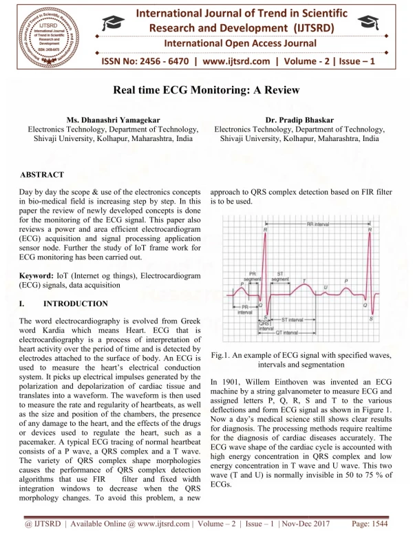
ECG interpretation Rate Rhythm Intervals QRS complexes ST segments & T waves
Normal Abnormal
ECG abnormalities Myocardial ischemia / infarction arrhythmias
Myocardial ischemia / infarction • ST depression (0.1mv) • ST elevation (0.2mv) • T wave inversion • Abnormal Q
Bradyarrhythmias Sinus Bradycardia
Bradyarrhythmias Junctional rhythm
Bradyarrhythmias 1st Degree AV block
Bradyarrhythmias: 2nd degree AVB Mobitz type I
Bradyarrhythmias: 2nd degree AVB Mobitz type II
Tachyarrhythmias: Premature complexes Atrial Premature Complexes
Tachyarrhythmias: Premature complexes Ventricular Premature Complexes
Tachyarrhythmias A.Fib
Tachyarrhythmias Atrial Flutter
Tachyarrhythmias PSVT
Tachyarrhythmias Ventricular Tachycardia
Tachyarrhythmias Torsades de pointes
Tachyarrhythmias Ventricular Fibrillation