Download
1 / 72
730 likes | 892 Views
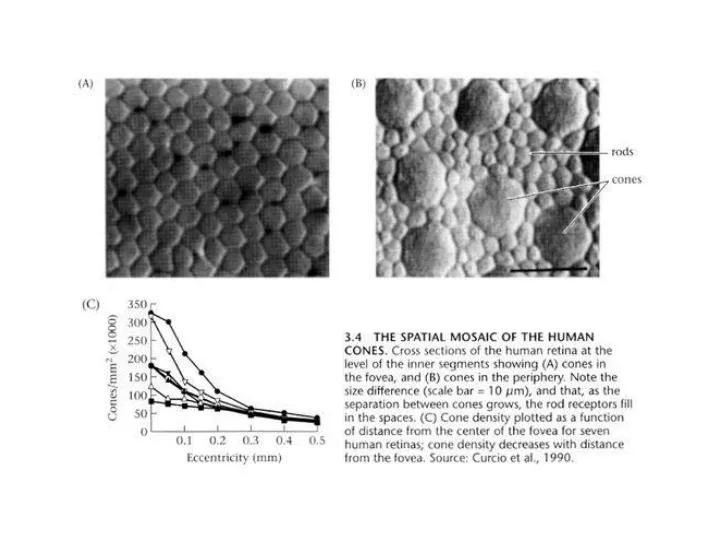
Foveal cones are about 2.4 μ m in diameter (0.7 min of arc) Peripheral cones are about 5.8 μ m in diameter (1.7 min of arc). Ganglion cells. Ganglion cells. Claim that ganglion cells are not photosensitive. (But, recall Pritchard.)

E N D
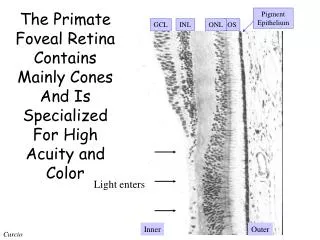
Foveal cones are about 2.4 μm in diameter (0.7 min of arc) • Peripheral cones are about 5.8 μm in diameter (1.7 min of arc)
Ganglion cells • Claim that ganglion cells are not photosensitive. (But, recall Pritchard.) • Ganglions cells fire action potentials, where photoreceptors use graded potentials.
Ganglion cells ~ 1.25 million ganglion cells 10-15 types of RGC ~ 100 million rods ~ 5 million cones Recall that, in photoreceptors, more light means less electrical current.
Ganglion cells are responsive only to limited areas of the visual field; they have limited receptive fields. • Ganglion cells can be ON-center with an OFF-surround. • Ganglion cells can be OFF-center with an ON-surround.
Lateral Inhibition • Stimuli in the surround suppresses activity in the center. • In other words, surround stimuli are antagonistic to center stimuli.
Ganglion cells with this concentric arrangement (i.e., ON-center, OFF-surround or OFF-center, ON-surround) found primarily in mammals with a fovea. Retinas lacking foveas, unlike most mammalian retinas, can process image motion and direction of motion.
How do photoreceptors, amacrine, bipolar, and horizontal cells work to make ganglion cells respond as they do? Blake & Sekuler are pretty sketchy on amacrine cells. Amacrine cells are inhibitory. Amacrine cells come in about 50 morphological types.
Horizontal cells Each horizontal cell connects to numerous photoreceptors. Each photoreceptor connects to numerous horizontal cells. Horizontal cells inhibit inactive photoreceptors more than active photoreceptors.
Wässl, H. (2004). Parallel Processing in the Mammalian Retina. Nature Reviews Neuroscience, 5, 1-11.
Bipolar cells • These contact photoreceptors. • Some bipolars: More current, less active. • Some bipolars: More current, more active. • More light, less current, more active. • Less light, more current, more active. • Inverting • Non-inverting
Some numerical details • In the fovea, the receptive field center of a retinal ganglion cell is connected to one cone cell. • In the fovea, the surround of retinal ganglion cell is connected to around 1-3 cone cells.
Receptive Field Size • The farther from fovea, the larger the field. • At each degree of eccentricity, there are “big field” and “small field” cells. The “big” fields are roughly three times the area of the “small” fields.
Three types of retinal ganglion cells • M cells (magnocellular) (large) • P cells (parvocellular) (small) • K cells (koniocellular) (very small) * Footnote 2, p. 82 has the terminology backward. M cells are parasol; P cells are midget.
M cells vs. P cells • M are bigger, hence conduct more quickly. • P cells are more numerous (80% of ganglion cells in primates are P cells) • For any degree of eccentricity, P cells have smaller receptive fields. • M cells are more responsive to luminance differences • M cells are faster responders than are P cells. • M cells are color indifferent; P cells are color selective.
Analysis of Perceptual Phenomena in Terms of Processes in Ganglion Cells • Hermann Grid Illusion • Mach bands
Questions about the Hermann Grid Illusion • Why are spots only at the intersection? • Why are there no spots in central vision?
Problems for the RGC Theory • Blake & Sekuler: • Illusion varies with orientation. • Illusion varies with spatial extent of grid • Illusion varies with regularity of grid
Problems for the RGC Theory Schiller & Carvey, 2005: • Illusion is perceived over a large range of sizes. • Illusion is reduced when the grid is rotated by 45º. • Illusion can be reduced by manipulations that do not alter the antagonistic/surround activation of retinal ganglion cells. • The ratio of the square size to the width of the intersecting bars is an important factor.
Problems for the RGC Theory Schiller & Carvey, 2005: • Enhancing center/surround antagonism at the intersections of bars does not enhance the illusory effect. • Varying contrast and color produces illusory effects not readily handled by the theory. • The spatial arrangement of RGC receptive fields is not what has been assumed by the theory.
Lightness contrast: Physically identical materials appear to differ in lightness. • Previous example • Lightness constancy: Physically distinct materials appear to be the same in lightness. • Text in bright sunlight and indoors
Does RGC activity explain lightness constancy? • Hans Wallach, (1963): An RGC will produce the same response to the same ratio of center-surround illumination.