Download
1 / 98
1.01k likes | 1.09k Views
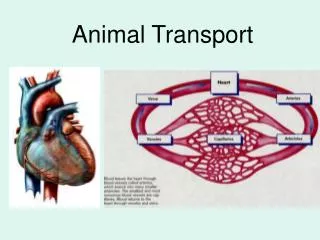
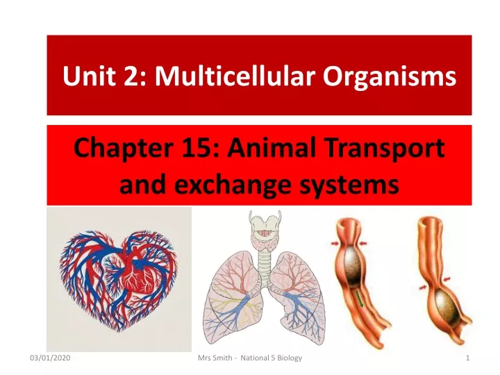
Unit 2: Multicellular Organisms. Chapter 15: Animal Transport and exchange systems. Unit 2: Multicellular Organisms. Chapter 15: Animal Transport and exchange systems. Lesson 1: Mammalian circulatory system: HEART. By the end of this chapter you should:

E N D
Unit 2: Multicellular Organisms Chapter 15: Animal Transport and exchange systems Mrs Smith - National 5 Biology
Unit 2: Multicellular Organisms Chapter 15: Animal Transport and exchange systems Lesson 1: Mammalian circulatory system: HEART Mrs Smith - National 5 Biology
By the end of this chapter you should: • Be able to demonstrate the pathway of oxygenated and deoxygenated blood through heart, lungs and body. • Describe the heart structure to include right and left atria and ventricles and location and function of valves (you DO NOT NEED valve names). Blood vessels to include: aorta, vena cava, pulmonary arteries and veins. Learning Intentions: Animal Transport Mrs Smith - National 5 Biology
Introduction • A large multicellular organism has a small surface area in relation to its volume. (See Torrance. ch14 – pg111). • Therefore it needs additional absorbing areas to take in oxygen and food. Mrs Smith - National 5 Biology
The need for additional absorbing areas in humans • Humans have for example • Alveoli in the lungs • Villi in the intestine These greatly increase the surface area for the absorption of oxygen and digested food respectively Mrs Smith - National 5 Biology
The need for a circulatory system • Once the essential substances have entered the animal’s body, they must be carried to all of it’s living cells at a faster rate than is possible by diffusion. • In mammals, this rapid transport of essential materials is achieved by an animal’s circulatory system. Mrs Smith - National 5 Biology
Mammalian circulatory system • The circulatory system consists of the • Heart (a muscular pump) • Blood vessels (a system of tubes) • These carry blood to all parts of the body. • Nutrients, oxygen, carbon dioxide and hormones are transported in the body. Mrs Smith - National 5 Biology
The Heart The heart is a muscular pump located in the centre of our chest. Its job is to pump blood all around the body. • Group Task – Study this diagram of a section through a human heart. • Try and work out – • How many chambers (large spaces) there are in the heart? • Why one side is coloured blue and one side coloured red? • Why one side has a thicker wall than the other side? Mrs Smith - National 5 Biology
The Heart - Chambers • The heart is divided into two separate sides. • Each side has 2 hollow chambers - an atrium and a ventricle. • The upper chambers are the right and left atria these collect blood. • The lower chambers are the right and left ventricles, these pump blood. • The wall of the heart is made of cardiac muscle. The diagram shows the 4 chambers viewed from the front of a person. The right hand side of a person is therefore on the left of the diagram and vice versa. Mrs Smith - National 5 Biology
The function of veins and arteries. We will look at their structure later! Veins carry blood into the heart while arteries carry blood away from the heart. The veins carry blood into the atria while the arteries carry blood away from the ventricles. vein Pulmonary vein artery Pulmonary Artery artery vein Aorta Vena cava Mrs Smith - National 5 Biology
Blood flow • This diagram shows the path taken by blood as it flows through the heart and its associated vessels. • Let’s look at it in more detail. Mrs Smith - National 5 Biology
Blood flow and associated vessels • Our blood flows through three different types of blood vessels is shown below. VEIN HEART ARTERY CAPILLARIES Mrs Smith - National 5 Biology
Flow of blood: Summary • The flow of blood from the LV back to the LV... As follows. • LV> aorta> body> vena cava> RA> RV> pulmonary artery> lungs> pulmonary vein> LA> LV. Pulmonary Artery Pulmonary vein Aorta Vena cava Mrs Smith - National 5 Biology
Blood flow – In more detail • Blood passes through the heart twice (each time it passes around the body also called double circulation). • The blood firstly is pumped to the lungs where it picks up oxygen, becoming oxygenated. • The blood is then pumped around the body where respiring cells remove the oxygen. This deoxygenates the blood. • The blood vessels that carry deoxygenated blood to the lungs are pulmonary arteries, while pulmonary veins return oxygenated blood to the heart. • The artery that carries oxygenated blood away from the heart and around the body is the aorta. The vena cava is the vein that returns deoxygenated blood to the heart from the body. Mrs Smith - National 5 Biology
Rob the Red Blood Cells Journey! Rob is a new red blood cell and needs direction about where he’s going and what will happen to him on his journey around the body. Can you help advise him? ? Mrs Smith - National 5 Biology
Review Questions • Where did Rob pick up Oxygen? • Where did Rob take it to? • Why did Rob change colour? • What things would be floating around Rob in the blood? • What other cells are there in the blood with Mrs Smith - National 5 Biology
Thickness of ventricle walls The muscle wall of the left ventricle is THICKER than the right ventricle. This is because the left ventricle has to pump blood right round the body, whereas the right ventricle only has to pump blood to the lungs. (which are right next to the heart). Mrs Smith - National 5 Biology
The Heart - Valves The heart has four valves in it. The job of the valves is to keep blood flowing in one direction through the heart. The valves stop the blood flowing backwards within the heart. Valve 4 Valve 3 Valve 2 Valve 1 Mrs Smith - National 5 Biology
The Heart – Valves 1 and 2 • Valves 1 and 2 are situated between the atria and the ventricles. • When they open, blood passes from the atria into the ventricles. • When the ventricles contract, the blood, under pressure closes valves 1 and 2. This prevents blood flowing back into the atria. Mrs Smith - National 5 Biology
The Heart – Valves 3 and 4 • Valves 3 and 4 are situated between the ventricles and the two arteries that leave the heart. • Once blood has been pumped through valves 3 and 4 they close, preventing backflow of blood from arteries into ventricles. • Blood is therefore only able to travel in one direction through the heart. Mrs Smith - National 5 Biology
Our heart beat is caused by the valve opening and closing Mrs Smith - National 5 Biology
Heart valve disease • If one or more valves are diseased or damaged, it can affect how blood flows through the heart in two ways: • If your valve does not open fully, it will obstruct the flow of blood. • If the valve does not close properly, it will allow blood to leak backwards. • People with heart valve disease may be advised to have surgery on your valve, which can greatly improve the symptoms and quality of life. Mrs Smith - National 5 Biology
Heart valve surgery There are two options for valve surgery: valve repair and valve replacement. • Valve repair is often used for mitral valves that become floppy and leak but are not seriously damaged. • Valve replacement is when the diseased valve is replaced with a new valve. The most common types of replacement valves are mechanical (artificial) valves or tissue (animal) valves. Mrs Smith - National 5 Biology
Consolidation exercise: The Heart Individual task - collect the diagram which represents a section through a human heart and stick it into the middle of a page in your jotter. • Now see if you can complete and label the diagram by - • naming the four chambers. • drawing and shading in the muscular walls of each ventricle. • indicating the positions of the four valves. • naming the four blood vessels associated with the heart. • drawing arrows to show the flow of blood through the heart. • colouring the “correct” side of the heart blue and the other side red. • indicating what the red and blue colours represent. Mrs Smith - National 5 Biology
The Heart: Watch this Watch the following video clip about the human heart. Heart : Twig Mrs Smith - National 5 Biology
Unit 2: Multicellular Organisms Chapter 15: Animal Transport and exchange systems Lesson 2: Mammalian circulatory system: BLOOD VESSELS Mrs Smith - National 5 Biology
By the end of this chapter you should: • Be able to compare the structure and function of arteries, veins and capillaries. • Specifically that • Arteries have thick, muscular walls, a narrow central channel and carry blood under high pressure away from the heart. • Veins carry blood under low pressure; have thinner walls and a wide channel. Veins contain valves to prevent backflow of blood and carry blood towards the heart. • Capillaries form networks at organs and tissues, are thin walled and have a large surface area, allowing exchange of materials. • Describe coronary arteries and their function. Learning Intentions: The vessels associated with the heart Mrs Smith - National 5 Biology
There are three types of blood vessels Mrs Smith - National 5 Biology
Artery Vein Task: Comparing Arteries and Veins • Group Task – • Study these diagrams of an artery and a vein. • Stick copies into your notes. • Describe threedifferences in the structure of arteries and veins. • Decide in which blood vessel the pressure will be highest. Give a reason for your choice. Mrs Smith - National 5 Biology
Vein Artery Blood Vessels ANSWER: Comparing Arteries and Veins Arteries have thick, muscular walls and a narrow central channel. Veins have thinner walls and a wide channel. They also contain valves to prevent the backflow of blood. Arteries carry blood at high pressure AWAY from the heart. Veins carry blood under low pressure back to the heart. • Individual Task – Study your wrists. • Are the blood vessels that you can see near the surface, arteries or veins? • Can you find your pulse in your wrist? What do you think your pulse actually is? Mrs Smith - National 5 Biology
What is a pulse? Each time the heart beats, blood is forced along the arteries at high pressure and this pressure wave can be felt as a pulse beat. Mrs Smith - National 5 Biology
Capillaries fun facts • There are an estimated 10 billion capillaries, measuring approximately 25,000 miles, in the average human body. • Each capillary has a length of about 1.1 millimeter. • Most capillaries are little more than a single cell layer thick. • Capillaries are the smallest and most numerous vessels in the body through which blood flows. • The thin capillary wall helps to increase the exchange of materials between cells in the tissue and the blood. • While a person is resting, approximately 5% of the blood circulating is in the capillaries, Mrs Smith - National 5 Biology
Capillary networks Mrs Smith - National 5 Biology
Capillaries • An artery divides into smaller vessels and finally into a dense network of tiny, thin walled capillaries. • Capillaries are the most numerous type of blood vessel in the body. They present a large surface area and are in close contact with all living cells in tissues and organs. This diagram shows the network of capillaries spreading through the skin in a fingertip. The larger blood vessels are small arteries which carry the blood to the capillaries. Individual Task – Press on the tip of your fingernail and watch what happens. Can you explain the change in colour that occurs. Mrs Smith - National 5 Biology
Capillaries cont • Capillaries are often referred to as exchange vessels since all exchanges of materials between blood and living tissue takes place through their thin walls (only one cell thick). • Capillaries unite to form larger vessels that converge to form veins. • The diagram shows a simplified version of the human circulatory system. Mrs Smith - National 5 Biology
TASK COPY: Capillaries Blood flows from arteries into capillaries and then back to veins. As the blood flows through capillaries substances are exchanged with the nearby body cells This diagram shows a capillary network in the inner lining of a cheek. Notice how all the cheek lining cells are very close to a blood capillary. • Group Task – Decide whether the following substances will be leaving or entering the blood at this capillary network. • Oxygen • Carbon dioxide • Glucose Mrs Smith - National 5 Biology
Task: Stick your diagram + complete the task below. Individual Task – Stick the diagram of a capillary network into your notes. Colour the blood vessels red and the cytoplasm of the body cells yellow. Individual Task - below your diagram draw one enlarged body cell with a capillary beside it. Individual Task – now draw arrows to represent the movement direction (diffusion) of 1) oxygen 2) carbon dioxide 3) glucose Mrs Smith - National 5 Biology
Blood Vessels: Summary: Watch this Watch the following video clip which contains information about blood vessels and the blood that flows around in them. Blood : Twig Mrs Smith - National 5 Biology
Coronary Artery This diagram shows the outside of the heart. Small arteries can be seen branching off the aorta. These are called coronary arteries and they are very important as they supply the muscles in the wall of the heart with oxygen. Mrs Smith - National 5 Biology
Coronary Heart Attack • The coronary arteries can become narrower as we get older. • A fatty diet, smoking and lack of exercise all contribute to this. • Eventually, a clot can block the artery leading to a heart attack. Muscle cells in the heart wall become starved of oxygen and die. The heart may stop contracting and unless the heart is quickly restarted the individual will die. Mrs Smith - National 5 Biology
Unit 2: Multicellular Organisms Chapter 15: Animal Transport and exchange systems Lesson 3: Mammalian circulatory system: BLOOD Mrs Smith - National 5 Biology
By the end of this chapter you should: • Be able to describe that mammals, nutrients, oxygen and carbon dioxide are transported in the blood. • Red blood cells are specialised by being biconcave in shape, having no nucleus and containing haemoglobin. This allows them to transport oxygen efficiently in the form of oxyhaemoglobin. Learning Intentions: Blood Mrs Smith - National 5 Biology
Remember blood is made of different cells Mrs Smith - National 5 Biology
Blood • Blood is a liquid tissue consisting of: • plasma • red blood cells • white blood cells • platelets • One of the functions of blood is to transport materials around the body. White blood cells and platelets are part of the body's immune system, but plasma and red blood cells are involved in transport. Mrs Smith - National 5 Biology
Task: Can you name three nutrients that are carried in the blood? • Group Task – Look at this diagram which represents the parts of blood. • Decide which part carries • oxygen • carbon dioxide • nutrients. • How many did you get correct? Mrs Smith - National 5 Biology
The Blood Nutrients such as glucose and amino acids dissolve in the fluid part of the blood – the plasma. Carbon dioxide gas is also carried around the body in blood plasma. Oxygen is transported around the body in red blood cells. Group Task – can you find out what white blood cells and platelets do? Mrs Smith - National 5 Biology
Plasma Plasma is a straw-coloured liquid. It transports dissolved substances around the body, including: • Hormones, (Remember hormones are chemical substances that help to regulate processes in the body)such as insulin which regulates the level of glucose in the blood. Oestrogen and progesterone are two hormones involved in the female menstrual cycle. • Nutrients, such as water, glucose, amino acids, minerals and vitamins • Waste substances, such as carbon dioxide and urea Mrs Smith - National 5 Biology
Microscopy - Seeing the composition of Blood This diagram shows a drop of blood as viewed under a microscope. The nuclei of the white blood cells have been stained purple to make them visible. Notice how few white cells there are compared to red cells. Mrs Smith - National 5 Biology
Why is blood is red? This diagram represents an image of blood magnified thousands of times using an electron microscope. The bright red colour of the red blood cells comes from the presence of a pigment called haemoglobin. Haem = Containing iron Mrs Smith - National 5 Biology