Download
1 / 18
180 likes | 181 Views
Learn about the complex process of muscle biopsy for diagnosing mitochondrial myopathies and the importance of proper planning and coordination between clinicians, pathologists, and technical staff.

E N D
http://www.mda.org/Publications/mitochondrial_myopathies.htmlhttp://www.mda.org/Publications/mitochondrial_myopathies.html
After the biopsy arrives in the pathology laboratory, it undergoes a complex series of studies. The pathologist uses knowledge of the clinical features to assist in interpretation of the constellation of pathologic findings in the biopsy and to help determine whether additional studies are warranted for a given patient. Therefore, muscle biopsy is somewhat complex in that an optimal outcome requires coordination of the clinician, surgical team, pathologist, and technical staff in the pathology laboratory. As muscle biopsies are often interpreted at specialized centers, a courier service also may need to be involved in the process; this is yet one more link in the chain from procedure to diagnosis. Unsuitable, suboptimal, or inadequate biopsy specimens usually can be attributed to lack of planning and forethought; no excuse exists for this situation. The single most important point to remember when contemplating muscle biopsy is to call the pathology laboratory in advance for advice on how to proceed.
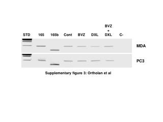
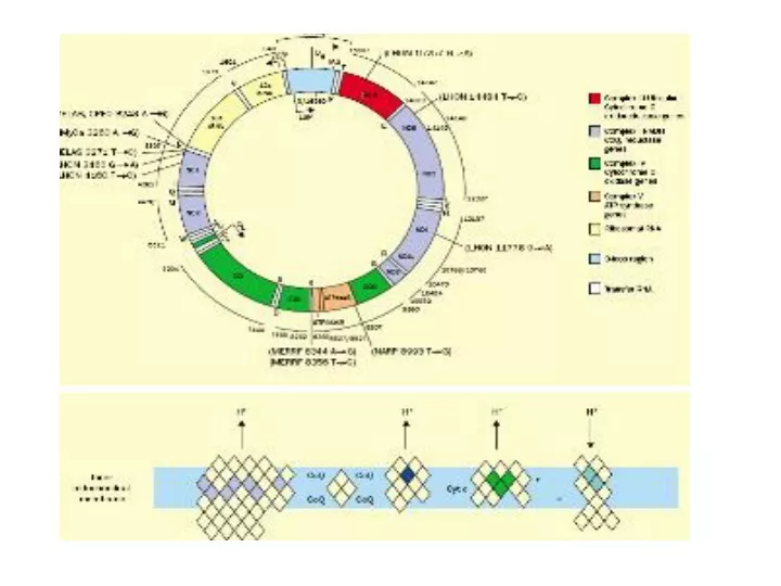
HUMAN SKELETAL BIOPSY FROM PATIENTS WITH DIFFERENT MITOCHONDRIAL MYOPATHIES my = myofibril
Xs normal skeletal (cytoplasmic and nuclear stain – brightfield microscopy)
TYPE II FIBER (fast twitch, mainly gycolytic) TYPE I FIBER (more sustained contraction , mainly oxidative phosphorylation) Xs normal skeletal muscle (NADH-tetrazolium stain LM)
http://www.pathology.vcu.edu/education/em/muscle.path.f.html