Download
1 / 1
10 likes | 138 Views
Charged Particle Therapy Treating cancer with protons and light ions. Cancer – a terrible disease

E N D
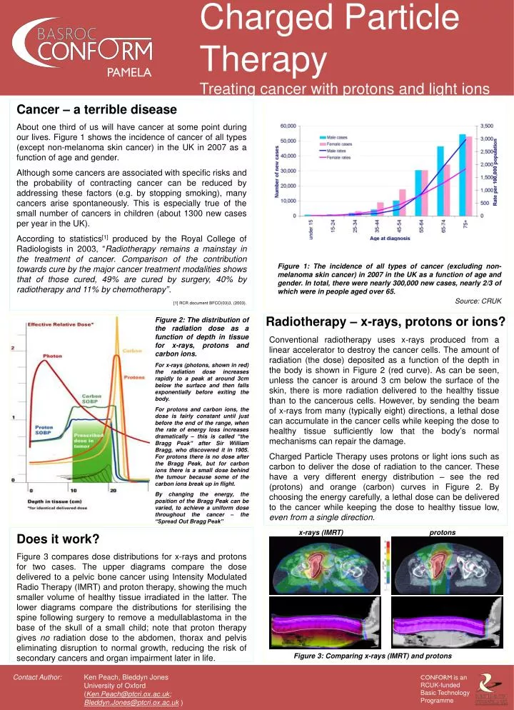
Charged Particle Therapy Treating cancer with protons and light ions Cancer – a terrible disease About one third of us will have cancer at some point during our lives. Figure 1 shows the incidence of cancer of all types (except non-melanoma skin cancer) in the UK in 2007 as a function of age and gender. Although some cancers are associated with specific risks and the probability of contracting cancer can be reduced by addressing these factors (e.g. by stopping smoking), many cancers arise spontaneously. This is especially true of the small number of cancers in children (about 1300 new cases per year in the UK). According to statistics[1] produced by the Royal College of Radiologists in 2003, “Radiotherapy remains a mainstay in the treatment of cancer. Comparison of the contribution towards cure by the major cancer treatment modalities shows that of those cured, 49% are cured by surgery, 40% by radiotherapy and 11% by chemotherapy”. [1] RCR document BFCO(03)3, (2003). Figure 1: The incidence of all types of cancer (excluding non-melanoma skin cancer) in 2007 in the UK as a function of age and gender. In total, there were nearly 300,000 new cases, nearly 2/3 of which were in people aged over 65. Source: CRUK Radiotherapy – x-rays, protons or ions? Figure 2: The distribution of the radiation dose as a function of depth in tissue for x-rays, protons and carbon ions. For x-rays (photons, shown in red) the radiation dose increases rapidly to a peak at around 3cm below the surface and then falls exponentially before exiting the body. For protons and carbon ions, the dose is fairly constant until just before the end of the range, when the rate of energy loss increases dramatically – this is called “the Bragg Peak” after Sir William Bragg, who discovered it in 1905. For protons there is no dose after the Bragg Peak, but for carbon ions there is a small dose behind the tumour because some of the carbon ions break up in flight. By changing the energy, the position of the Bragg Peak can be varied, to achieve a uniform dose throughout the cancer – the “Spread Out Bragg Peak” Conventional radiotherapy uses x-rays produced from a linear accelerator to destroy the cancer cells. The amount of radiation (the dose) deposited as a function of the depth in the body is shown in Figure 2 (red curve). As can be seen, unless the cancer is around 3 cm below the surface of the skin, there is more radiation delivered to the healthy tissue than to the cancerous cells. However, by sending the beam of x-rays from many (typically eight) directions, a lethal dose can accumulate in the cancer cells while keeping the dose to healthy tissue sufficiently low that the body’s normal mechanisms can repair the damage. Charged Particle Therapy uses protons or light ions such as carbon to deliver the dose of radiation to the cancer. These have a very different energy distribution – see the red (protons) and orange (carbon) curves in Figure 2. By choosing the energy carefully, a lethal dose can be delivered to the cancer while keeping the dose to healthy tissue low, even from a single direction. x-rays (IMRT) protons Does it work? Figure 3 compares dose distributions for x-rays and protons for two cases. The upper diagrams compare the dose delivered to a pelvic bone cancer using Intensity Modulated Radio Therapy (IMRT) and proton therapy, showing the much smaller volume of healthy tissue irradiated in the latter. The lower diagrams compare the distributions for sterilising the spine following surgery to remove a medullablastoma in the base of the skull of a small child; note that proton therapy gives no radiation dose to the abdomen, thorax and pelvis eliminating disruption to normal growth, reducing the risk of secondary cancers and organ impairment later in life. Figure 3: Comparing x-rays (IMRT) and protons Ken Peach, Bleddyn Jones University of Oxford (Ken.Peach@ptcri.ox.ac.uk; Bleddyn.Jones@ptcri.ox.ac.uk)