Download
1 / 39
420 likes | 838 Views
The Meninges and Blood Vessels of Brain and Spinal Cord, and the Cerebrospinal Fluid. The Meninges of Brain and Spinal Cord. The spinal cord and brain are surrounded by three membranes, the meninges. Named from the outside inward they are the dura mater , arachnoid , and pia mater . .

E N D
The Meninges and Blood Vessels of Brain and Spinal Cord, and the Cerebrospinal Fluid
The Meninges of Brain and Spinal Cord • The spinal cord and brain are surrounded by three membranes, the meninges. Named from the outside inward they are the dura mater, arachnoid, and pia mater.
The meninges of spinal cord • Spinal dura mater • Spinal arachnoid mater • Spinal pia mater
Spinal dura mater Characters • Above, attached to circumference of foramen magnum • Below, becomes thinner at level of S2, invests filum terminale to attach at back of coccyx
Epidural space • Position: lies between spinal dura mater and periosteum of vertebral canal • Contents: a quantity of loose connective tissue, fat, lymphatic vessels and vertebral venous plexus, the spinal nerves on each side pass through the epidural space which is applicable for block anesthesia Subdural space
Spinal arachnoid mater Characters • A thin, delicate, tubular membran loosely investing spinal cord • Above, it is continuous with cerebral arachnoid mater
Subarachnoid space • Position: lies between pia and arachnoid maters containing cerebrospinal fluid • Terminal cistern : the largest part of subarachnoid space extending from termination of spinal cord to level of S2, where it is occupied by nerves of cauda equina, so it is the best site for a lumbar puncture
Spinal pia mater • A delicate vascular membrane that closely invests the spinal cord • Denticulate ligament: consist of 21 pairs triangular ligaments extending from spinal cord on each side between anterior and posterior roots of spinal nerves to spinal dura mate; these ligaments help to fix position of spinal cord. • Filum terminale: an extension of pia beyond conus medullaris
The Meninges of Brain • Cerebral dural mater • Cerebral arachnoid mater • Cerebral pia mater
Cerebral dural mater Characters • A thick and dense inelastic membrane that composed of two layers, an inner or meningeal and outer or endosteal • It is in loose contact with calvaria, and most strongly adherent to base of skull
Four septa • Cerebral falx • Tentorium of cerebellum - in front there is a gap, the tentorial incisure, for passage of midbrain • Cerebellar falx • Diaphragma sellae
Sinuses of duramater • Superior sagittal sinus • Inferior sagittal sinus • Straight sinus • Confluence of sinus
Transverse sinus • Sigmoid sinus • Superior petrosal sinuses • inferior petrosal sinuses
Cavernous sinus • Position: lies on each side of sella turcica • Relations of cavernous sinus: • Internal carotid artery and abducent nerve run through the sinus • Oculomotor and trochlear nerves and ophthalmic and maxillary divisions of trigeminal nerve lie in the lateral wall of the sinus
The flowing of the blood in dural sinus Sup. sagittal sinus Transverse sinus Inf. sagittal sinus Straight sinus Confluence of sinus Sup. petrosal sinus Sigmoid sinus Cavernous sinus Inf. petrosal sinus Internal jugular vein
Cerebral arachnoid mater • Characters: a delicate membrane covering brain loosely, passing over sulci and entering only cerebral longitudinal and transverse fissures
Arachnoid granulations -project into sinuses of dura mater, serve as sites where cerebrospinal fluid diffuses into bloodstream
Subarachnoid cisterns • Cerebellomedullary cistern • Interpeduncular cistern • Pontine cistern • Superior cistern
Cerebral pia mater • Closely invests brain surface, in some areas the pia invaginates into ventricles to take part in the formation of choroids plexus
Circulation of cerebrospinal fluid (CSF) • Cerebrospinal fluid is a clear colorless fluid, which acts as a transport medium for nutrients provides a protective fluid cushion for the central nervous system. • Production: produced by the choroids plexuses within the lateral, third and fourth ventricles
Circulation of cerebrospinal fluid interventricular foramina third ventricle CSF drains from lateral ventricle mesencephalic aqueduct median and two lateral apertures fourth ventricle arachnoid granulations vein subarachnoid space superior sagittal sinus
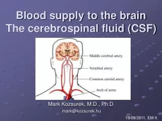
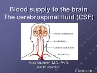
Arteries of brain Two sources • Internal carotid artery: supplies anterior 2/3 of cerebral hemisphere and parts of diencephalon • Vertebral artery: supplies postterior 1/3 of cerebral hemisphere and parts of diencephalon, brain stem and cerebellum
Internal carotid artery • Anterior cerebral artery • Middle cerebral artery • Anterior choroidal artery • Posterior communicating artery
Anterior cerebral artery • Joined the fellow of the opposite side by the anterior communicating artery • Cortical branches: supply all medial surface of the cerebral cortex as far back as the parietooccipital sulcus and superior border of the suprolateral surface of the cerebral hemisphere • Central branches: supply caudate nucleus, anterior part of lentiform nucleus and anterior limb of internal capsule
Middle cerebral artery • Cortical branches: supply most of superolateral surface of cerebral hemisphere and insular lobe • Central branches: supply lentiform and caudate nuclei, genu and posterior limb of internal capsule (lenticulostriate artery)
Anterior choroidal artery: passes backward, enters inferior horn of lateral ventricle, and ends in choroid plexus. It supplies lateral geniculate body, posterior limb of internal capsule, middle 3/5 of crus cerebri,and globus pallidus • Posterior communicating artery: runs backward to join posterior cerebral artery
Vertebral artery • Cranial branche • Anterior and posterior spinal arteries • Posterior inferior cerebellar artery • Branches of basilar artery • Anterior inferior cerebral artery • Labyrinthine artery • Pontine arteries • Superior cerebellar artery • Posterior cerebral artery
Posterior cerebral artery • Cortical branches: supply medial and inferior surfaces of temporal lobe and occipital lobe • Central branches: supply dorsal thalamus, medial and lateral geniculate bodies, hypothalamus and subthalamus
Cerebral arterial circle ( circle of Willis ) • Formation: formed by anterior communicating artery, both anterior cerebral arteries, internal carotid arteries, posterior communicating arteries, and posterior cerebral arteries • Position: lies on sella turcica around optic chiasma, tuber cinereum and mamillary bodies
Area of oxygendepriveds brain Blockage Thrombus Plaque
Microaneurysm Lenticulostriate arteries Subarachnoid hemorrhage
Intracerebral hemorhage Arteriovenous malformation
Veins of brain Superficial cerebral veins • Drain blood from cortex and subcortical medullary substance and empty into adjacent sinuses of dura mater
Veins of brain • Deep cerebral veins: drain deeper parts of hemispheres, basal nuclei, internal capsule, diencephalon and choroid plexus, ultimately form great cerebral vein which enter straight sinus
Blood vessels of spinal cord Arteries of spinal cord • Two sources • Anterior and posterior spinal arteries • Branches of segmental arteries: radicular arteries of posterior intercostals arteries, lumbar arteries, and lateral sacral arteries • Damage area: T1~T4,ventral part of L1 • vascular ring ( vasocorona )
Blood vessels of spinal cord Spinal veins: drain into internal vertebral venous plexus