Download
1 / 59
630 likes | 1.13k Views
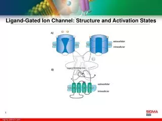
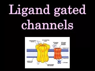
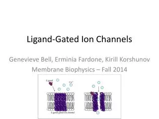
Ligand-Gated Ion Channels. Jony mallik M.Pharm. Contents. General Information Ligand-Gated Ion Channels The Acetylcholine Receptor Neurotransmitters Molecular Diversity and its Control Toxin targets. Channel Families. Voltage-gated Extracellular ligand-gated Intracellular ligand-gated

E N D
Ligand-Gated Ion Channels Jony mallik M.Pharm
Contents • General Information • Ligand-Gated Ion Channels • The Acetylcholine Receptor • Neurotransmitters • Molecular Diversity and its Control • Toxin targets
Channel Families • Voltage-gated • Extracellular ligand-gated • Intracellular ligand-gated • Inward rectifier • Intercellular • Other
Typical Ion Channels with Known Structure: Acetylcholine receptor M2 transmembrane segment K+ channel (KCSA) • Types of ion channels: • Simple pores (GA, GAP junctions) • Substrate gated channels (Nicotinic receptor) • Voltage-gated channels (K-channels) • Pumps (ATP-synthase, K+,Na+-ATPase)
What are the Biochemical Changes that Lead to Channel Gating (Opening or Closing)? Gating involves some type of conformational change in the protein, but other than that there are few definitive answers to the question. However, there are several general proposed models for gating.
Types of Biochemical Mechanisms that Open and Close Channels • Conformational change occurs in a discrete area of the channel, leading to it opening. • The entire channel changes conformation (e.g., electrical synapses). • Ball-and-chain – type mechanism. • Nt or hormone binding causes the channel to open.
Types of Biochemical Mechanisms that Open and Close Channels (Cont’d) • Nt or hormone binding to receptor causes a 2nd messenger to activate a protein kinase that phosphorylates a channel and thus opens it. • Changes in membrane potential. • Membrane deformation (e.g., mechanical pressure). • Selectivity by charge (i.e., positively lined pore allows anions through; negatively lined pore allows cations through).
Extracellular ligand-gated • nicotinic ACh (muscle): 2 (embryonic), 2 (adult) • nicotinic ACh (neuronal): (2-10), (2-4) • glutamate: NMDA, kainate, AMPA • P2X (ATP) • 5-HT3 • GABAA: (1-6), (1-4), (1-4), , , (1-3) • Glycine
Intracellular ligand-gated • leukotriene C4-gated Ca2+ • ryanodine receptor Ca2+ • IP3-gated Ca2+ • IP4-gated Ca2+ • Ca2+-gated K+ • Ca2+-gated non-selective cation • Ca2+-gated Cl– • cAMP cation • cGMP cation • cAMP chloride • ATP Cl– • volume-regulated Cl– • arachidonic acid-activated K+ • Na+-gated K+
G-protein linked receptors coupled to ion channels • Acetylcholine (muscarinic) • Adenosine & adenine nucleotides • Adrenaline & noradrenaline • Angiotensin • Bombesin • Bradykinin • Calcitonin • Cannabinoid • Chemokine • Cholecystokinin & gastrin • Dopamine • Endothelin • Galinin • GABA (GABAB) • Glutamate (quisqualate) • Histamine • 5-Hydroxytryptamine (1,2) • Leukotriene • Melatonin • Neuropeptide Y • Neurotensin • Odorant peptides • Opioid peptides • Platelet-activating factor • Prostanoid • Protease-activated • Tachykinins • Taste receptors • VIP • Vasopressin and oxytocin
Gated Ion Channels • Another type of membrane transport • Pores in the membrane that open and close in a regulated manner and allow passage of ions -“Dispose” of the gradients • Passive transporters -Ions flow from high to low concentration -No energy is used -If there is no gradient ions will not flow
Gated Ion Channels • Small highly selective pores in the cell membrane • Move ions or H2O • Fast rate of transport 107 ions x s-1 • Transport is always down the gradient • Cannot be coupled to an energy source
Ion channels are everywhere • Channels are present in almost every cell • Functions -Transport of ions and H2O -Regulation of electrical potential across the membrane -Signaling
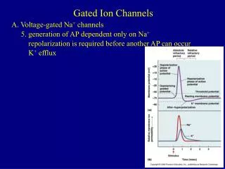
Gating mechanisms • Two discrete states ;open (conducting) closed (nonconducting) • Some channels have also inactivated state (open but nonconducting) • Part of the channel structure or external particle blocks otherwise open channel
What gates ion channels? • Non gated - always open • Gated Voltage across the cell membrane Ligand Mechanical stimulus, heat (thermal fluctuations)
Gating mechanisms • Conformational changes in channel protein are responsible for opening and closing of the pore -3D conformational shape is determined by atomic, electric, and hydrophobic forces • Energy to switch the channel protein from one conformational shape to another comes from the gating source
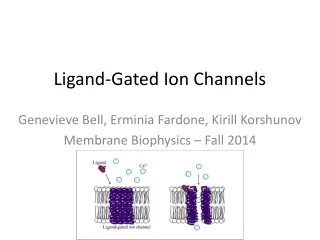
Ligand gated channels • Glutamate receptors • Nicotinic acetylcholine receptor • Vanilloid receptor family (TRPV) Ion Flow = Current = Neurotransmitter
Ligand gated ion channels • Gated by ligands present outside of the cell • In fact they are receptors • All of them are nonselective cation channels • Mediate effects of neurotransmitters
Acetylcholine Receptor • consists of a pentamer of protein subunits, with two binding sites for acetylcholine, which, when bound, alter the receptor's configuration and cause an internal pore to open. • This pore allows Na+ ions to flow down their electrochemical gradient into the cell. a b ACh g (ore) d ACh
Structure of the AChR at 4.6Å Miyazawa et al, (1999) (left figure was modified)
filled site in ACh binding protein Brejc, 2001
Channel Gating Mechanisms AChR: Proposed gating mechanism (Unwin, 1995) Open Closed
The ACh receptor also responds to nicotine, and so is called the “nicotinic” acetylcholine receptor -nAChR
the resting (closed) ion channel to acetylcholine (ACh)producesthe excited (open) state. Longer exposure leads to desensitization and channel closure. Acetylcholine binding sites Continued excitation Na+, Ca2+ ACh Outside Inside Resting (gate closed) Desensitized (gate closed) Excited (gate open) ACh
•Synaptic transmission throughout the nervous system is predominantly Chemical •At the chemical level, the key players include integral membrane proteins that control signaling
Neurotransmission is fast and precise Action Potential opens voltage gated Ca2+ channels Ca2+ enters the terminal. Ca2+ initiates vesicular release of neurotransmitter Note that nerve transmission of the AP involves BOTH VG ion channels AND LG ion channels.
Mechanism of Transmitter release Reserve vesicles are outside the active zone. Synapsins tethers vesicles to the cytoskeleton Ca+2 activates Ca2+/calmodulin dependent protein kinase which phosphorylates synapsin I and frees the vesicles.
Molecular Diversity and its Control Genes and Gene Expression: • Sets of gene families encoding an ion channel receptor correspond to the sets of protein subunits of the same functional class: e.g., There are gene families each for the major α, β, and γ subunits for GABAA receptors, and for the GluR1-4 (AMPA), Glur5-7 (kainate), KA-1 and KA-2 (kainate), and NR2A-D (NMDA) glu receptor subunits.
Molecular Diversity and its Control Genes and Gene Expression: • Generally, the genes for each receptor class are scattered over many chromosomes, with occasional clusters; e.g., human GABAA receptor α1, α6, β2, and γ2 genes are all close together at q31-35 on chromosome 5. What is this called…?
Molecular Diversity and its Control Genes and Gene Expression: • Introns – There does not seem to be any consistency in the lengths of the introns and exons and, therefore, the genes, of various receptor genes both between and within a species. e.g., the gene encoding the mouse GABAA receptor δ subunit is ~ 14 kb long, but the β1 subunit gene, which has the same intron-exon structure, is 65 kb long.
Molecular Diversity and its Control Genes and Gene Expression: • Transcriptional Control Most attention, to date, given to: • Timing of gene expression during development. • Mechanism of expression of gene expression to neurons and types of neurons. • Regulation of receptor gene expression during synapse formation. However, relatively little is known about the mechanism by which gene expression responds to environ signals impinging on the individual neuron.
Molecular Diversity and its Control Genes and Gene Expression: • Multiprotein transcriptional complex consisting of RNA Pol II and a plethora of ancillary factors. • TATA box? Not all genes have them! • Some use other initiator elements. • Simple binding to TATA box insufficient to transcribe a gene to satisfy physiol levels of txn. • Need additional seq-specific interactions of various txn factors with cis-acting enhancer and silencer elements.
Molecular Diversity and its Control Genes and Gene Expression: • Much evidence for the role of silencing in neurons acting at 2 levels: • Global silencing of neuronal genes in non-neuronal cell types. • Silencing at the fine-tuning level to restrict expression of neuronal genes to a subset of neurons. What is the most important example of #2?
Molecular Diversity and its Control Genes and Gene Expression: • The GluR subunit genes have been the most heavily investigated. • Have no TATA nor CAAT start sites. • GC-rich with multiple txn start sites within a CpG island. • Promotors contain overlapping Sp1 and GSG recog sites near the major txn start sites and an NSR silencer. • NSR sequence in the NR1 and GluR2 genes has a small modulatory effect with respect to neuronal specificity of expression.
Molecular Diversity and its Control Genes and Gene Expression: • The GluR2 NSR sequence is a site of mediation of the stimulatory effects on gene expression of the signaling pathways initiated by the neurotrophic factor, GDNF and BDNF. • Ligand-switching-induced changes in gene expression: occurs a lot during development; e.g., replacement of the nAchR fetal γ subunits by functionally homologous ε subunits in myocytes. e.g., fetal expression of only GlyR α2 subunits to adult expression of only GlyR α1 and α3 subunits in the s.c. underlies the differential sensitivities to strychnine affinities for the GlyR subtypes. e.g., NR2B to NR2C NMDAR subunits in cereb gran cells ~ 2 wks after birth.
Molecular Diversity and its Control Alternative Splicing: • Not very widespread among the RNA transcripts of LG ion channel receptors. • Where it does occur, often entails little more than a difference in a short aa sequence that may regulate postranslational modification processes, such as phosphorylation and glycosylation sites. • Alt Splicing also produces variants of the same receptor (e.g., AMPA) subunits.
Molecular Diversity and its Control Alternative Splicing: e.g., Each of the 4 AMPAR subunits occur in 2 alternatively spliced variants, called flip and flop. Correspond to the alternative inclusion of either of 2 adjacent exons (exons 14 and 15 in the GluR2 gene). Functional difference: the flip forms of most subunits desensitize more slowly and to a lesser degree than the flop forms - distinction that is easily displayed probed with various agents. • NR1 subunit of the NMDARs has 3 alt spliced exons 8 splice variants variations of the C-term exons different receptor clustering properties in the membrane (e.g., only NR1 variants with the C2” cassette interact with the PSD) and Zn2+ and H+ sensitivities.
Molecular Diversity and its Control RNA Editing: • As ---- oxidative deaminated inosine • Inosine bp like G Δ the codon different aa in the translated protein. • Carried out by 2 dsRNA A deaminases, ADAR1 and ADAR2. • ADAR1 and ADAR2 depend on the formation of ds structures involving intronic sequences, which bp with the exonic sequences to be edited. • Other protein factors prob participate as well, because some cells express ADARs, but cannot edit.
Molecular Diversity and its ControlRNA Editing GluR Q/R Editing GluR R/G Editing GluR2, 3, 4 undergo editing at the R/G site in exon 13, just N-term to the flip flop region of alt splicing. Reduces sensitization and accel recovery. • $ RNA editing sites: GluR2, 5, 6: replacement of a gln codon (CAG) by an arg codon (CIG=CGG) and insertion of arg (R) into the TM domain • Occurs in M2 has low Ca2+ permeability, low single-channel conductance and linear rectifying properties.
Molecular Diversity and its Control Translational Control: • Not exactly known how widespread this mech is among LG ion channel subunits. • Thus, should not make inferences about the levels of protein subunits from the mRNAs. • In many GluR subunits and NR subunits, removal of (some e.g., 15 bp) the 5’UTR that is involved in the putative stem-loop structure results in signif disinihibition of translation. • Translational suppression of gluR2 mRNA largely due to broad region containing a repeat sequence near the 5’ end of the mRNA, which may affect various txn start sites differentially.
Molecular Diversity and its Control Post-translational Modification: • Extensively phosphorylated and glycosylated. • Kinases known to phosphorylate LCICs: PKA, CaMKII, PKC. • Phosphorylation by these kinases affect the function of LGICs: i. PKA phos AMPARs incr channel open time or the P(O) state. ii. CaMKII phos AMPARs corr with incr synaptic responses (synaptic plasticity). iii. Phosphorylation of receptors is corr with activity.
Molecular Diversity and its Control Post-translational Modification: • Phosphorylation (and dephosphorylation) efficiency dependent on proximity to of LGICs to kinases and phosphatases in the high-[protein] milieu. E.g., For gluRs, PSD-95/SAP97 required for clustering. E.g.,For AKAPs (A-kinase anchoring protein) anchor kinases and phosphatases to the complex. E.g., RACK-1 stimulates PKC to phosphorylate GABAAR at the β subunits by binding these subunits to at a site different from PKC.
Molecular Diversity and its Control Post-translational Modification: • ~ 5-10% of a subunit MW can be glycosylation. • Oligomannosidic glycans + complex oligosaccharides. • Incr the efficiency of receptor assembly and cell-surface density of GABAARs , but not essential for these processes. • But is strongly required for ER exit of assembled GlyR and nAchR. • Appears that glycosylation has a similar quality control over all receptors. • Apart from this, there does not seem to be a clear general role for glycosylation of LGICs.
Molecular Diversity and its Control Receptor Assembly and Trafficking: • Peptide synthesis. • Folding. • Post-translational modifications. • Insertion into ER. • Strict selectivity is required so that only certain combinations of subunits (and the corrects ones!) are oligomerized and targeted to the plasma membrane via the Golgi.