Download
1 / 27
270 likes | 356 Views
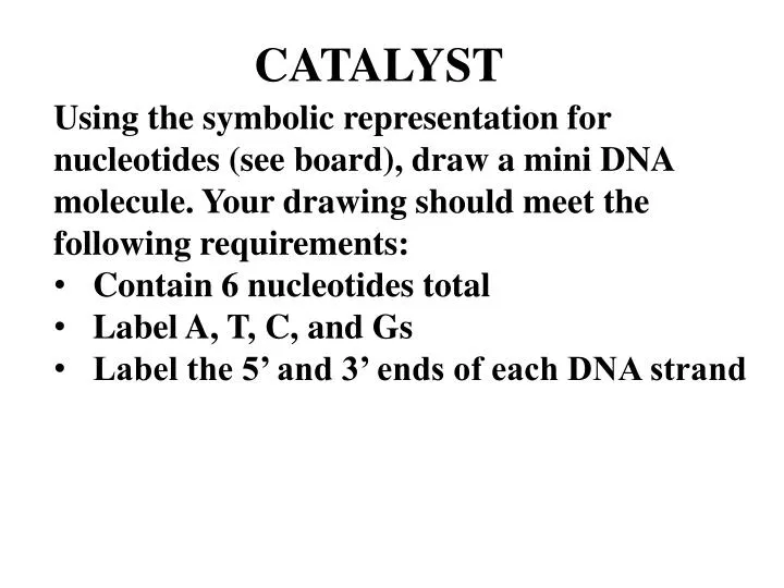
Using the symbolic representation for nucleotides (see board), draw a mini DNA molecule. Your drawing should meet the following requirements: Contain 6 nucleotides total Label A, T, C, and Gs Label the 5’ and 3’ ends of each DNA strand. CATALYST. TODAY: PROTEINS!.

E N D
Using the symbolic representation for nucleotides (see board), draw a mini DNA molecule. Your drawing should meet the following requirements: Contain 6 nucleotides total Label A, T, C, and Gs Label the 5’ and 3’ ends of each DNA strand CATALYST
The Building Blocks of Proteins: AMINO ACIDS!
Practice Peptide Bonding! • Draw two glycine amino acid molecules side by side (glycine’s R group is just “H” • Circle what atoms must be removed in order for a peptide bond to form • Draw the dipeptide that is formed
Primary Structure Secondary Structure! Hydrogen bonding!
Tertiary Structure Quaternary Structure (in some proteins) Hemoglobin’s Chemical Formula: C3032H4816O872N780S8Fe4
Change in primary structure = Change in function! Mutated Cells
Change in structure = Change in function!
Change in structure = Change in function!
Levels of Protein Structure – Group Demo
Levels of Protein Structure • In your group of 3 – 4 people… • 1) Create a song / rap • 2) Include all 4 levels of protein structure and the types of bonds / attractive forces involved in each level
From 20 building blocks… A HUGE DIVERSITY OF STRUCTURE!
Two Main Categories: 1) FIBROUS • Structure: • Water insoluble • Very tough • Can be supple or stretchy • Parrallel chains in long fibers or sheets
Two Main Categories: 1) FIBROUS • Function: • Structural support - collagen in connective tissue • Contractile – actin and myosin in muscles
Two Main Categories: 2) GLOBULAR • Structure: • Water soluble • Tertiary / quaternary structure critical to function
Two Main Categories: 2) GLOBULAR • Function: Protective (antibodies)
Two Main Categories: 2) GLOBULAR • Function: Transport (aquaporins, hemoglobin)
Two Main Categories: 2) GLOBULAR • Function: Catalytic (enzymes)
Two Main Categories: 2) GLOBULAR • Function: Regulatory (hormone receptors)
When Protein’s Go Awry = PRIONS! Normal Misfolded (Prion) • Scrapie • Kuru • Mad Cow
Practice Free Response 10 points One of the unifying themes in biology is that function follows structure. Explain how the structure of proteins determines their function by responding to the following: Describe the structure of an amino acid (3) Outline the formation of a protein from its primary structure to its quaternary structure (4 points) Discuss the relationship between protein structure and function in fibrous and globular proteins (4)