Download
1 / 30
310 likes | 881 Views
BLOOD. CIRCULATORY SYSTEM 3. BLOOD VESSELS. Arteries - conduct blood away from the heart Arterioles - little arteries Capillaries - concerned with exchange between blood and tissues Venules - little veins Veins - conduct blood to the heart. CIRCULATORY ROUTES.

E N D
BLOOD CIRCULATORY SYSTEM 3
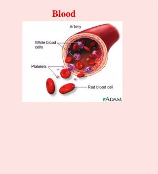
BLOOD VESSELS • Arteries - conduct blood away from the heart • Arterioles - little arteries • Capillaries - concerned with exchange between blood and tissues • Venules - little veins • Veins - conduct blood to the heart
CIRCULATORY ROUTES 1. Heart arteries arterioles capillaries venules veins heart 2.Through a portal system- blood flows through two consecutive capillary networks before entering the heart 3.Through arteriovenous shunt - artery flows directly into vein 4.Through an anastomosis (point where two blood vessels merge)
PORTAL SYSTEMS • Hypothalamo-hypophyseal portal system between hypothalamus & anterior pituitary • Renal portal system in kidneys • Hepatic portal system between intestines and liver
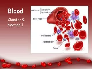
COMPONENTS OF VESSEL WALLS • Tunica externa - outermost layer -loose connective tissue • Tunica media - middle layer - smooth muscle, collagen, some elastic • Tunica intima (interna) - innermost layer of simple squamous endothelium on a basement membrane and a layer of fibrous tissue
The Vessel Wall Copyright © The McGraw-Hill Companies, Inc. Permission required for reproduction or display. Conducting (large) artery Large vein Lumen Lumen Tunica interna: Tunica interna: Endothelium Endothelium Basement membrane Basement membrane Tunica media Tunica media Tunica externa Tunica externa Vasa vasorum Vasa vasorum Nerve Nerve Inferior vena cava Medium vein Distributing (medium) artery Aorta Tunica interna: Tunica interna: Endothelium Endothelium Basement membrane Basement membrane Internal elastic lamina Valve Tunica media External elastic lamina Tunica media Tunica externa Tunica externa Direction of blood flow Arteriole Venule Tunica interna: Tunica interna: Endothelium Endothelium Basement membrane Basement membrane Tunica media Tunica media Tunica externa Tunica externa Endothelium Basement membrane Capillary
CAPILLARIES They are extremely narrow and have thin, permeable walls 1. Continuouscapillaries: occur in most tissues Pericytes with contractile protein wrap around the capillaries and regulate blood flow 2. Fenestrated capillaries: found in organs that require rapid absorption or filtration - kidneys and small intestine Endothelial cells have numerous holes calledfiltration pores (fenestrations) 3. Sinusoids (discontinuous capillaries): in liver, bone marrow, spleen Irregular blood-filled spaces with large fenestrations Allow proteins (albumin), clotting factors, and new blood cells to enter the circulation
Continuous Capillary Copyright © The McGraw-Hill Companies, Inc. Permission required for reproduction or display. Pericyte Basal lamina Intercellular cleft Pinocytotic vesicle Endothelial cell Erythrocyte Tight junction
Fenestrated Capillary Copyright © The McGraw-Hill Companies, Inc. Permission required for reproduction or display. Endothelial cells Nonfenestrated area Erythrocyte Filtration pores (fenestrations) Basal lamina Intercellular cleft 400 µm (a) (b) b: Courtesy of S. McNutt
Sinusoid in Liver Copyright © The McGraw-Hill Companies, Inc. Permission required for reproduction or display. Macrophage Endothelial cells Erythrocytes in sinusoid Liver cell (hepatocyte) Microvilli Sinusoid
REGULATION OF BLOOD PRESSURE AND FLOW 1. Local control 2. Neural control 3. Hormonal control
LOCAL CONTROL OF BLOOD PRESSURE AND FLOW • Autoregulation - waste accumulation leads to vasodilation • Vasoactive chemicals e.g. histamine and bradykinin stimulate vasomotion • Reactive hyperemia - blood supply cut off then restored • Angiogenesis - growth of new vessels - controlled by growth factors and inhibitors
NEURAL CONTROL OF BLOOD PRESSURE AND FLOW • Involves vasomotor center of medulla oblongata • - sympathetic control stimulates most vessels to constrict, but dilates vessels in skeletal and cardiac muscle • - integrates three autonomic reflexes • baroreflexes • chemoreflexes • medullary ischemic reflex
Use Slide show View Please note that due to differing operating systems, some animations will not appear until the presentation is viewed in Presentation Mode (Slide Show view). You may see blank slides in the “Normal” or “Slide Sorter” views. All animations will appear after viewing in Presentation Mode and playing each animation. Most animations will require the latest version of the Flash Player, which is available at http://get.adobe.com/flashplayer.
HORMONAL CONTROL OF BLOOD PRESSURE AND FLOW • Angiotensinogen (prohormone produced by liver) Renin (kidney enzyme - low BP) • Angiotensin I ACE (angiotensin-converting enzyme in lungs) ACE inhibitors block this enzyme lowering BP • Angiotensin II • very potent vasoconstrictor
HORMONAL CONTROL OF BLOOD PRESSURE AND FLOW • Aldosterone - promotes Na+ and water retention by the kidneys - increases blood volume and pressure • Atrial natriuretic factor - increases urinary sodium excretion - generalized vasodilation • ADH - promotes water retention • Epinephrine and norepinephrine - vasoconstriction ofmost blood vessels, but vasodilation ofskeletal and cardiac muscle blood vessels
Major Systemic Arteries • Supplies oxygen and nutrients to all organs
Arterial Pressure Points • Some major arteries close to body surface allow palpation for pulse and serve as pressure points to reduce arterial bleeding.
Major Branches of the Aorta • Right & left coronary arteries supplying heart branch from ascending aorta • Branches from aortic arch: • brachiocephalic trunk - branches into right common carotid a. supplying right side of head, and right subclavian a. supplying right shoulder & upper limb • left common carotid a. supplying left side of head • left subclavian a. supplying shoulder and upper limb • Descending aorta is thoracic aorta above diaphragm and abdominal aorta below diaphragm
Major Branches of the Aorta Copyright © The McGraw-Hill Companies, Inc. Permission required for reproduction or display. L. common carotid a. R. common carotid a. R. subclavian a. L. subclavian a. Brachiocephalic trunk Aortic arch Ascending aorta Descending aorta, thoracic (posterior to heart) Diaphragm Aortic hiatus Descending aorta, abdominal
Arteries of the Head and Neck Copyright © The McGraw-Hill Companies, Inc. Permission required for reproduction or display. Supraorbital a. Superficial temporal a. Ophthalmic a. Posterior auricular a. Occipital a. Maxillary a. Facial a. Internal carotid a. External carotid a. Lingual a. Carotid sinus Superior thyroid a. Vertebral a. Thyroid gland Common carotid a. Thyrocervical trunk Costocervical trunk Subclavian a. Axillary a. Brachiocephalic trunk (a) Lateral view
Arterial Supply of the Brain • Paired vertebral arteries combine to form the basilar artery on the pons • Circle of Willis on base of brain is formed from anastomosis of basilar & paired internal carotid arteries • Supplies brain, internal ear and orbital structures • anterior, middle & posterior cerebral • superior, anterior & posterior cerebellar
Arteries of the Upper Limb • Subclavian changes names as it passes to different regions • Subclavian to axillary to brachial to radial & ulnar • Brachial is used for measuring blood pressure. • Radial artery is used for measuring pulse.
Arteries of the Thorax • Thoracic aorta supplies viscera & body wall • bronchial, esophageal and mediastinal branches • posterior intercostal and phrenic arteries • Internal thoracic, anterior intercostal & pericardiophrenic arise from subclavian artery
Arteries of the Lower Limb • Branches to the lower limb arise from external iliac branch of the common iliac artery
Major Systemic Veins • Deep veins run parallel to arteries while superficial veins have many anastomoses • Deep veins run parallel to arteries while superficial veins have many anastomoses
Deep Veins of Head and Neck • Large, thin-walled dural sinuses form in between layers of dura mater and drain blood into internal jugular vein
Superficial Veins of Head & Neck • Branches of internal and external jugular veins drain the external structures of the head • Upper limb is drained by subclavian vein
Veins of the Head and Neck Internal jugular vein receives most of the blood from the brain Copyright © The McGraw-Hill Companies, Inc. Permission required for reproduction or display. Superior ophthalmic v . Superficial temporal v . Occipital v. Facial v . Vertebral v. External jugular v . Superior thyroid v . Thyroid gland Internal jugular v . Subclavian v . Axillary v. Brachiocephalic v . (c) Superficial veins of the head and neck
Veins of Hepatic Portal System • Drains blood from viscera (stomach, spleen, pancreas and intestines) to liver