Download
1 / 25
250 likes | 428 Views
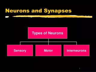
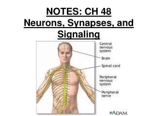
Ch. 48 Neurons, Synapses, and Signaling. A central nervous system (CNS) and a peripheral nervous system (PNS) process information in three stages: sensory input, integration, and motor output to effector cells.

E N D
A central nervous system (CNS) and a peripheral nervous system (PNS) process information in three stages: sensory input, integration, and motor output to effector cells.
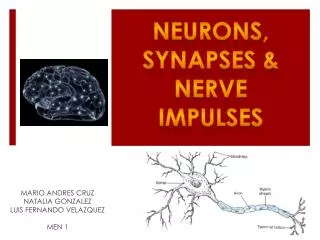
Most neurons have branched dendrites that receive signals from other neurons and an axon that transmits signals to other cells at synapses. • Neurons rely on glia for functions that include nourishment, insulation, and regulation.
48.2 Ion pumps and ion channels • Ionic gradients generate a voltage difference, or membrane potential, across the plasma membrane of cells. • The concentration of Na+ is higher outside than inside; the reverse is true for K+.
In resting neurons, the plasma membrane has many open potassium channels but few open sodium channels. • Diffusion of ions, principally K+, through channels generates a resting potential, with the inside more negative than the outside.
48.3 Action potentials are the signals conducted by axons • Neurons have gated ion channels that open or close in response to stimuli, leading to changes in the membrane potential. • An increase in the magnitude of the membrane potential is a hyperpolarization; a decrease is a depolarization.
Changes in membrane potential that vary continuously with the strength of a stimulus are known as graded potentials.
An action potential is a brief, all or none depolarization of a neuron’s plasma membrane. • When a graded depolarization brings the membrane potential to the threshold, many voltage-gated ion channels open, triggering an inflow of Na+ that rapidly brings the membrane potential to a positive value.
A negative membrane potential is restored by the inactivation of sodium channels and by the opening of many voltage-gated potassium channels, which increases K+ outflow. • A refractory period follows, corresponding to the interval when the sodium channels are inactivated.
A nerve impulse travels from the axon hillock to the synaptic terminals by propagation of a series of action potentials along the axon. • The speed of conduction increases with the diameter of the axon and, in many vertebrate axons, along with myelination.
Action potentials in myelinated axons jump between the nodes of Ranvier, a process called saltatory conduction.
48.4 Neurons communicate with other cells at synapses • In an electrical synapse, electrical current flows directly from one cell to another. • In a chemical synapse, depolarization causes synaptic vesicles to fuse with the terminal membrane and release neurotransmitter into the synaptic cleft.
At many synapses, the neurotransmitter binds to ligand-gated ion channels in the postsynaptic membrane, producing an excitatory or inhibitory postsynaptic potential (EPSP or IPSP). • The neurotransmitter then diffuses out of the cleft, is taken up by surrounding cells, or is degraded by enzymes.
Temporal and special summation at the axon hillock determines whether a neuron generates an action potential.
Different receptors for the same neurotransmitter produce different effects. • Some neurotransmitter receptors activate signal transduction pathways, which can produce long-lasting changes in postsynaptic cells.
Major neurotransmitters include acetylcholine; the amino acids, GABA, glutamate, and glycine; biogenic amines; neuropeptides; and gases such as NO.