Download
1 / 112
1.15k likes | 1.28k Views
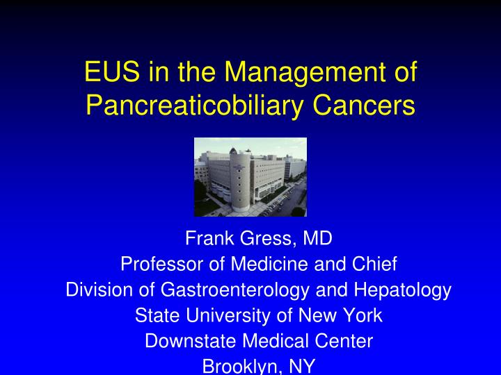
EUS in the Management of Pancreaticobiliary Cancers. Frank Gress, MD Professor of Medicine and Chief Division of Gastroenterology and Hepatology State University of New York Downstate Medical Center Brooklyn, NY.

E N D
EUS in the Management of Pancreaticobiliary Cancers Frank Gress, MD Professor of Medicine and Chief Division of Gastroenterology and Hepatology State University of New York Downstate Medical Center Brooklyn, NY
"EUS for the Diagnosis, Staging FNA and Celiac Neurolysis of Pancreatic Cancer”
Pancreatic Adenocarcinoma • The fourth leading cause of cancer-related death in the U.S. • At diagnosis only ~15% of patients are candidates for curative surgery • Five-year survival following a Whipple procedure was only 25% for node-negative tumors and 10% for node-positive tumors Ahmad et al. Long term survival after pancreatic resection for pancreatic adenocarcinoma. The American Journal of Gastroenterol 2001;96(9):2609-15
Pancreatic Cancer • Late presentation, aggressive nature and lack of effective therapies all contribute to the poor prognosis • Early detection is crucial to improve the overall prognosis • Accurate Staging is vital for selecting the subset of patients who have potentially resectable tumors
Common Indications for EUS GI Tumor Staging • Esophageal Cancer • Gastric Cancer • Rectal Cancer • Ampullary Cancer • Pancreatic Cancer
Cancer Staging EUS Staging Accuracy Compared to Path IndicationnT stageN stage Esophageal CA 739 85% 79% Gastric CA 1163 78% 73% Pancreatic CA 155 90% 78% Ampullary CA 94 86% 72% Rectal CA 19 84% 84%
Clinical Applications for EUS Pancreatic and Biliary Disease • Tumor Staging • Localization of Endocrine Tumors • Detecting Choledocholithiasis • Detecting Chronic Pancreatitis
EUS Indications for Staging • Pancreatic Masses • Adenocarcinoma • Other malignancies/metastases • Bile duct cancer (cholangiocarcinoma)
Clinical Applications for EUS CurrentIndications Pancreatic and Biliary Malignancies 1) Tumor staging primarily based on ability to assess for vascular invasion 2) Localization of Endocrine Tumors 3) Ability to sample lesions for diagnosis with >85% accuracy
EUS Stations for Staging Pancreatic Tumors Transducer Major Structures Location identified with EUS Gastric Body Confluence, Body/Tail of Pancreas, PD, Celiac Axis, Splenic vessels, SMA Gastric Antrum Gallbladder,Liver,Pancreas Duodenum Bulb Head of Pancreas, CBD, PD 2nd Portion Head of Pancreas, SMA/SMV, Aorta, PD, Ampulla, Liver
EUS Staging of Pancreatic Cancer TNM Classification T Staging is based on tumor size, depth of invasion and infiltration into major vessels N Staging assesses for nodal involvement M Staging denotes the absence/presence distant metastasis (EUS can detect hepatic metastasis)
EUS Detection Rates of Pancreatic Tumors Sensitivity (%) Specificity (%) PPV(%) NPV(%) Accuracy (%) Rosch, 1991 99 100 100 97 76 Snady, 1992 85 80 89 73 83 Yasuda, 1993 - - - - 100 Muller, 1994 94 100 - - 96 Gress, 199793 100 - - - Baron, 1997 95 88 95 88 - Legmann, 1998 100 93 - - - Akahoshi, 1998 89 97 94 93 94 Totals 95 94 95 88 90
Pancreatic Cancer Staging by EUS Pooled Data • T staging accuracy ranges from 78 to 94% • T staging accuracy is higher in patients with advanced lesions (T3 and T4) • Vascular invasion accuracy was 82 to 93% • N staging accuracy ranges from 64 to 82%
Diagnosis by EUS • EUS provides improved imaging of small tumors not seen with other imaging modalities • The detection of pancreatic tumors < 3 cm in diameter was higher for EUS: • EUS (100%) • TUS (57%) • CT (68%) Rosch, et al. Endoscopic ultrasound in small pancreatic tumors. Z Gastroenterol 1991;29:110-5.
Diagnosis by EUS • The detection of pancreatic tumors < 2cm in diameter was higher for EUS: • EUS (100%) • ERCP (57%) • TUS (29%) • CT (29%) • Angiography (14%) Yasuda K, et al. The diagnosis of pancreatic cancer by endoscopic ultrasonography. Gastrointest Endosc 1998;34:1.
EUS Staging Lower T and N staging accuracy has also been described: • 89 patients with pancreatic cancer had EUS staging compared to surgery • Overall accuracy for T staging was 69% and for N staging was 54% • Only 46% of tumors designated by EUS as resectable actually were at laparotomy Ahmad,et al Gastrointest Endo 2000;52:46
Pancreatic Cancer • Best modality for small lesions • Diagnostic imaging and fine needle aspiration during single procedure • Evaluate for chronic pancreatitis if not tumor found • All pancreatic cancer has a dismal prognosis
Liver • EUS provides excellent imaging of the liver particularly the left lobe of the liver and some portions of the right lobe • The left lobe is best seen from the gastric body and fundus • The right lobe is best imaged from the antrum and duodenum
Clinical Utility of EUS FNA for Diagnosing Liver lesions • Sensitivity of EUS-FNA for the diagnosis of malignancy ranged from 82 to 94% • When compared with benign lesions, EUS features predictive of malignant hepatic masses were the presence of regular outer margins (60% vs 27%; p = 0.02) and the detection of two or more lesions (38% vs 9%; p = 0.03). [DeWitt J et al. Am J Gastroenterol. 2003 Sep;98(9):1976-81]
EUS IndicationsCancer Staging • Ampullary Most accurate locoregional staging • Rectal Most accurate locoregional staging • Other e.g. duodenal tumors, adenomas
Limitations of EUS • Factors influencing EUS staging accuracy: • Experience level of endosonographer • Imaging artifacts/Normal variants/Chronic Pancreatitis • Distinguishing vascular compression from tumor infiltration can be difficult in larger tumors • Accuracy for detecting invasion into the SMA and SMV is lower than that for PV or SV
EUS versus Helical CT Contrast enhanced helical CT has been compared to EUS for detecting pancreatic tumors, predicting resectability and determining vascular invasion Leggmann, et al; 1998 Midwinter, et al; 1999 Mertz, et al; 2000 Tierney, et al; 2002
EUS versus Helical CT Pooled Data Accuracy 4 Studies n=164 EUS CT Detecting pancreatic tumors 97% 73% Predicting resectability 91% 83% Determining vascular invasion 91% 64% Hunt GC, et al. Gastrointest Endosc 2002;55:232.
EUS versus Helical CT • Several features of the individual studies may account for the disparity in the conclusions: - Differences in Gold Standard - Differences in Helical CT Technique - Number of patients with advanced disease
EUS versus Multidetector CT • Prospective study comparing EUS and Multidetector CT for detecting and staging pancreatic cancer 120 patients with known pancreatic cancer EUS was: 98% sensitive for tumor detection (86% for CT) 67% for tumor staging accuracy (41% for CT) 44% for nodal staging accuracy (47% for CT) DeWitt J, et al. Comparison of EUS and Multidetector CT for detecting and staging pancreatic cancer. Annals of Internal Med 2004;141:753-63
EUS versus Helical CT Conclusions • EUS and Helical CT are complementary for staging pancreatic cancer. • EUS is a more accurate modality for T staging and predicting vascular invasion and CT is better for detecting distant metastasis.
EUS and Cancer Diagnosis • Controversial whether pre-operative diagnosis is necessary • Direct to resection when clinical suspicion is high vs. • Pre-operative tissue diagnosis
EUS-FNA of Pancreatic Lesions Not all pancreatic masses are cancer Differential Diagnosis • Adenocarcinoma • Neuroendocrine tumor • Lymphoma • Chronic pancreatitis
Normal Pancreas EG-3630UR
Normal Pancreas GF-UM130 EG-3630UR
Chronic Pancreatitis EG-3630UR
EUS-guided Fine Needle Aspiration • Percutaneous or CT-guided biopsy has been the traditional approach for establishing the diagnosis of pancreatic cancer • EUS FNA was introduced ~10 years ago • The main advantage of EUS guided FNA biopsy is its ability to obtain tissue sampling of any suspicious mass found during EUS evaluation.
Diagnostic Characteristics of EUS FNA for Pancreatic Mass Lesions n Sensitivity (%) Specificity (%) Accuracy (%) Giovannini[Endoscopy 1995;27(2)] 43 75 100 79 Cahn[AJS 1996;172(5)] 50 88 100 87 Bhutani[Endoscopy;1997;29(9)] 47 64 100 72 Chang[GIE;1997;45(5)] 44 92 100 95 Erickson[AFP 1997;55(6)] 28 -- -- 96 Faigel[JClinOnc1997;15(4)] 45 72 100 75 Gress[GIE1997;45(3)] 121 80 100 85 Wiersema[Gastro 1997;112(4)] 124 87 100 88 Binmoeller[GIE1998;47(2)] 58 76 100 92 560 81% 100% 86%