Download

1 / 38
380 likes | 557 Views
University of Pittsburgh Senior Design - BioE1161. OCT Penlight. Sara Hanrahan Brock Nichol Gina Rodriguez Areej Sajjad. Outline. Background Objectives Product design specifications OCT Penlight design Results Budget Validation & verification Future work. What is Glaucoma?.

E N D
University of Pittsburgh Senior Design - BioE1161 OCT Penlight Sara Hanrahan Brock Nichol Gina Rodriguez Areej Sajjad
Outline • Background • Objectives • Product design specifications • OCT Penlight design • Results • Budget • Validation & verification • Future work
What is Glaucoma? • Damage to the optic nerve that may lead to vision loss or complete blindness • 66.8 million people with visual impairment • 6.7 million suffering from blindness [1] [1] http://www.ahaf.org/glaucoma/about/glabout.htm - Accessed December 4, 2007
Possible Cause of Glaucoma • Schlemm’s canal clogged, preventing aqueous humor from escaping • High intraocular pressure (IOP) • Cassin, B. and Solomon, S. Dictionary of Eye Terminology. Gainsville, Florida: Triad Publishing Company, 1990.
Canaloplasty: A Promising Solution • Expand Schlemm’s canal to relieve IOP • New, safe, and minimally invasive technique (do not have to enter anterior chamber) • Biggest challenge: surgeon has to find Schlemm’s canal
Market Size • In 1999, 400,320 glaucoma surgeries were conducted in the US [2] • These patients could benefit from the OCT Penlight • There are approximately 7,500 hospitals in the US [3] [2] Strutten, Walt, J Glaucoma. 2004 Jun;13(3):221-6. [3]http://www.census.gov/PressRelease/www/releases/archives/facts_for_features_special_editions/004491.html
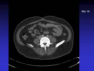
Optical Coherence Tomography (OCT) • Acquires a cross-sectional image of the eye • Penetrates about 2 mm into tissue • High resolution imaging technology • Currently used for diagnostic purposes OCT Handheld
Real-Time Tomographic Reflection (RTTR) Stetten, G., Chib, V., Hildebrand, D., Bursee, J. “Real Time Tomographic Reflection: Phantoms for Calibration and Biopsy.” IEEE/ACM International Symposiium on Augmented Reality. 2001 Oct:11-19. New York City.
Objectives • Long term goal: • Locate Schlemm’s canal with more ease during surgery • Short term goal: • Place a 30 gauge needle into a goat eye guided by the OCT penlight
OCT Penlight Components Software Mechanical Component Calibration Arm
Product Design Specifications • Place virtual image accurately (100 µm tolerance) • Allow a sufficient working area • Weigh less than 2 lbs • Clamp onto a 3 cm diameter lens • Software and hardware capable of acquiring image from OCT device
Mechanical Design • Virtual image plane must be placed along OCT scanning beam • Must be structurally stable • Must be fixed display mirror virtual image
Handheld OCT Patient OCT Software Display Signal Analysis Computer VGA Signal Splitter VGA Capture Device OCT Penlight Display OCT Penlight Computer
Handheld OCT Patient OCT Software Display Signal Analysis Computer VGA Signal Splitter VGA Capture Device OCT Penlight Display OCT Penlight Computer
Handheld OCT Patient OCT Software Display Signal Analysis Computer VGA Signal Splitter VGA Capture Device OCT Penlight Display OCT Penlight Computer
Handheld OCT Patient OCT Software Display Signal Analysis Computer VGA Signal Splitter VGA Capture Device OCT Penlight Display OCT Penlight Computer
Handheld OCT Patient OCT Software Display Signal Analysis Computer VGA Signal Splitter VGA Capture Device OCT Penlight Display OCT Penlight Computer
Handheld OCT Patient OCT Software Display Signal Analysis Computer VGA Signal Splitter VGA Capture Device OCT Penlight Display OCT Penlight Computer
Handheld OCT Patient OCT Software Display Signal Analysis Computer VGA Signal Splitter VGA Capture Device OCT Penlight Display OCT Penlight Computer
Handheld OCT Patient OCT Software Display Signal Analysis Computer VGA Signal Splitter VGA Capture Device OCT Penlight Display OCT Penlight Computer
Software Crop Display Display our software using OpenGL + GLUT Scale X Rotate Scale Y Translate Capture Screen
Calibration • Purpose • Guide user in positioning the virtual image to its accurate location on the scanning plane • Process • 3 pins within scanning range • Pins scanned and image displayed • User aligns virtual image • Easily detached 4.5 mm 2 mm
Prototype Fabrication • Stereolithography (SLA) • Lightweight • Cost-efficient • Sufficiently sturdy • Readily available
Final Product A: Flexible arm B: OCT Handheld Device C: Lens of OCT D: OLED Display E: Half-Silvered Mirror F: Calibration Arm
Displaying the Virtual Image mirror
Validation & Verification • Allow a sufficient working area • Approved by an experienced ophthalmologist • Weigh less than 2 lbs • Using a scale, we weighed entire device • Clamp onto a 3 cm diameter lens • OCT Penlight was able to clamp onto lens and maintain position for at least two hours
Validation & Verification • Software and hardware capable of acquiring image from OCT device • Run the software and see if image on monitor is visible on the Penlight display • Placement of virtual image must be accurate (100 µm tolerance) • Examine calibration process through a microscope • Objective must be met • Guide user in penetrating a goat eye with a 30 gauge needle using OCT Penlight
Future Work • Use a higher quality microscope to obtain clearer pictures of the virtual image • Attach OCT Penlight directly to surgical microscope • Automate calibration • Test Penlight on live specimens • Make software faster • More efficient video capture
Acknowledgements • Sources of funding • Generous gift of Drs. Hal Wrigley and Linda Baker • Department of Bioengineering • Mentors • Dr. George Stetten • Larry Kagemann • Additional help • Andy Holmes • Gaurav Shukla • Dr. John Galeotti • Dr. Bing Wu • Dr. Gadi Wollstein