Download
1 / 40
410 likes | 1.01k Views
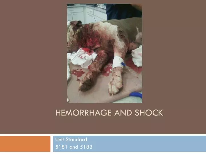
Hemorrhage and shock. Unit Standard 5181 and 5183. Haemorrhage/Bleeding. Haemorrhage Acute bleeding (sudden trauma or injury) Chronic bleeding (small amounts of blood being lost over period of time) What are some examples?. Haemorrhage.

E N D
Hemorrhage and shock Unit Standard 5181 and 5183
Haemorrhage/Bleeding • Haemorrhage • Acute bleeding (sudden trauma or injury) • Chronic bleeding (small amounts of blood being lost over period of time) • What are some examples?
Haemorrhage • What is the difference between arterial, venous and capillary bleeding? • Arterial bleeding spurts as the blood in the vessels is under pressure which rises and falls with each heart beat. This is the most serious form of haemorrhage; the blood is bright red. • Venous bleeding occurs as a steady flow of darker blood. • Capillary bleeding is a steady ooze and has multiple pinpoint sources from damaged tissue. This is bright red blood.
Haemorrhage • Questions for students to answer – • What do we mean when we talk about internal and external bleeding? • What is secondary bleeding? • Why does haemorrhage tend to stop of its own accord?
Haemorrhage • Questions for students to answer – • What disorders might be present if bleeding will not stop? • How would you go about stopping bleeding – consider internal and external?
Haemorrhage • Control must be achieved as soon as possible • You will need to assess the efficacy of the method used for control of bleeding and adjust if needed • 4 main methods of haemorrhage control • Direct Digital Pressure • Pressure Bandage • Pressure Points • Tourniquet • Use anything you have on hand – i.e. t-shirt, hat, jacket. • Advisable to have someone restraining when applying haemorrhage control, as can be painful
Haemorrhage • It is important not to ‘pat’ / ‘dab’ the wound as this may promote bleeding – instead apply steady pressure continuously. • Keep the pressure on the wound from 10 seconds to 30 minutes or until a vet can see the animal (depending on severity of bleeding). • If, after the pressure has been removed, the wound starts to bleed again, then reapply pressure. • Extra pressure or bandaging may be necessary for arterial bleeding.
Pressure Bandage • These are applied to extremities such as limbs • Direct pressure is applied to a wound by providing plenty of padding material and a firmly applied external bandage • If the wound is on the limb the entire distal limb (down to toes) should be bandaged to prevent swelling of the extremities • If a foreign body or fractured bone is protruding from the wound a ring pad should be placed first, then the limb bandaged.
Pressure Bandage Layer 1 • Layer 1 – contact with the wound • Should be non-stick • Jelonet – paraffin gauze • good non stick product, but has been demonstrated to slow healing • Telfa or Melolin Pads • non stick, used on wounds that are not exuding much or actively bleeding • Tape • Adhesive, so not appropriate for the wound but can be used to help secure bandage (stirrups)
Padding - Layer 2 • Cotton wool • especially good for Robert Jones type dressing where need a thick bandage that has a splinting effect • Animal lintex • used more commonly in Large Animals - has a cover over it that draws exudate away from the wound. Used in Poultice type dressings • Softban • A lighter padding layer, good for paw dressing and light bandages where splinting is not required. It is more a protective cover.
Layer 3 Holding it all together • Vetwrap • comes in lots of lovely colours – the owners love it especially when you do the bandage in local rugby colours!! Self adhesive, easy to apply, but does not stick to the animals coat so it is easier for the animal to remove • Elastoplast • usually put some strips of this over the vet wrap to give some waterproofing to the toe area (note it is not very waterproof, only more than vetwrap) and also a layer at the top to stick it to the coat – keeps the dressing on more successfully.
Direct Digital Pressure • Clean Fingers are applied directly to bleeding vessel • Finger and thumb are placed on either side and gently pinching the wound edges together to prevent further haemorrhage. • Ensure the wound is free from foreign bodies • Note - do not remove foreign bodies when you cannot feel the full extent of them • Care should be taken where fractures are present to avoid further displacement of bone fragments
Pressure Points • Pressure is applied on an artery (passing over a bone) to the body extremities. • The supply of blood to forelimbs, hind limbs or tail can be temporarily constricted. • The pressure required must be sufficient to prevent the flow of blood through the artery. It does not affect venous bleeding and is only a temporary measure.
Pressure Points • The pressure points are: • Brachial Artery • can be found on the distal third of the humerus, down the medial shaft. This will arrest haemorrhage below the elbow • Femoral artery • medial aspect of the femur, on the inner thigh (where the pulse is found). Pressure here will arrest haemorrhage to the stifle • Coccygeal artery • on the underside of the tail base. Pressure here will arrest haemorrhage to the rest of the tail.
Pressure Points • Brachial • Femoral
Tourniquets • These are used to cut off the blood supply to tissues below the application site. • Great care should be taken with a tourniquet • Avoid use if at all possible • Timing is vital for the survival of the tissues • A tourniquet should never be left on for more than 10 minutes, after which it must be taken off for 1-2 minutes to allow blood flow back into the tissue • It is only a temporary method until can get a dressing on. • Be aware of risks (loss of limb) involved with ANY use of a tourniquet.
Tourniquets • There are many forms available • Flat elastic band with fastening clip • Strong Bandage or material • Rubber tubing • Narrow Belt • DO NOT use string or rope as they cause severe tissue damage • Fix the tourniquet firmly around the limb/tail • A few inches above the wound and apply enough pressure to stop the haemorrhage, by adjusting the lip. • With improvised tourniquets, tie firmly onto the skin and tie a stick or rod over the knot and gradually twist to tighten
Haemorrhage Control • Once the haemorrhage is controlled • Observe and check the patient regularly, including dressings. • Make sure the patient is comfortable as possible • Try to estimate blood loss • Await or seek qualified help.
Revision • What is the difference between arterial, venous and capillary bleeding? • Arterialbleeding spurts; the blood is bright red. • Venousbleeding occurs as a steady flow of darker blood. • Capillarybleeding is a steady ooze and has multiple pinpoint sources from damaged tissue. This is bright red blood.
Revision • List the 4 main methods you can use to control haemorrhage • Direct Digital Pressure • Pressure Bandage • Pressure Points • Tourniquet
Revision • What is the maximum length of time you would leave a tourniquet on? Why? • 10 minutes maximum to prevent the tissue from dying
Shock • Shock • Circulatory failure in which cardiac output cannot meet the body’s oxygen and nutrient requirements. Removal of wastes is also inadequate, and will result in toxicity.
Classifications of Shock • Pending • the history of the patient is known and shock is expected to follow, but clinical signs are not yet evident. Interception at this point will have a favourable effect in preventing deterioration of the patient.
Classifications of Shock • Established • Shock is already established. The patient is showing clinical signs of shock and treatment is essential. With effective treatment the results should be favourable, as long as no further complications arise.
Classifications of Shock • Irreversible • Shock is irreversible, the patient has been suffering from shock for a considerable time, and despite treatment, the patient will likely deteriorate further and/or die.
Types of Shock • Hypovolemic shock • Loss of blood, plasma, water, and/or electrolytes resulting in inadequate blood volume • Cardiogenic shock • failure of the heart to maintain adequate blood circulation
Types of Shock • Vasogenic shock • Loss of tone in blood vessels, in conditions such as: • Endotoxic shock (septic) • Neurogenic shock (sudden loss of the autonomic nervous system) • Anaphylaxis (allergic reaction) • All these result in overall increase in the volume of blood vessels, and a decrease in the effective blood volume – even though the actual volume may not have changed. • Traumatic Shock • The body’s response to severe trauma is to shut down the non-essential circulation (hands and feet for example).
Signs of Shock (NOT cats) • Weak rapid pulse • Increased heart rate • Pale and/or dry mucous membranes • Slow CRT • Rapid, shallow respiration • Cold extremities • Subnormal rectal temperature • Decreased level of consciousness • Reduced urine output • Dilated pupils • Poor skin turgor • Weakness and depression
Signs of Shock - Cats • Weak slow pulse • Decreased heart rate • Pale and/or dry mucous membranes • Slow CRT • May have altered respiration • Cold extremities • Subnormal rectal temperature • Decreased level of consciousness • Reduced urine output • Dilated pupils • Poor skin turgor • Weakness and depression
Prevention and Treatment of Shock • Control and prevent further haemorrhage • Provide oxygen therapy • Via face mask, nasal tube or oxygen cage. • Provide intravenous access • For the infusion of I/V fluids ieHartmans and/or for emergency drugs. (under vet supervision) • Provide pain control
Prevention and Treatment of Shock • Restore body temperature • Provide insulation to the patient with towels, blankets, etc, by wrapping them in it. Hot water bottles, heat pads are acceptable as long as they are covered, and constantly checked. • Maintain constant observation • The patient may deteriorate rapidly, and immediate actions will need to be taken, so constant checking, observing and recording of details is essential. • Provide a comfortable kennel • It is essential that the patient is comfortable to avoid further deterioration. Quiet, warm surroundings, with subtle lighting will aid recovery.
First Aid Equipment for Shock • Out in the field • Blankets and towels • Wound dressing & bandaging equip • Thermometer • Stethoscope • Scissors • Back at the Clinic • Oxygen supply • Warm, darkened kennel • Pre-warmed I/V fluids • Infusion set • I/V catheter • Drugs for shock (as per practice policy) • Record Sheet • Plus everything you have in the field