Download
1 / 51
510 likes | 519 Views
Explore the vast and diverse world of the animal kingdom, with over 1.3 million identified species. Learn about their unique characteristics, nutritional modes, cell structure and specialization, reproduction and development, and the evolution of animals over millions of years. Discover the different body plans and symmetry found in animals.

E N D
Overview: Welcome to Your Kingdom • The animal kingdom extends far beyond humans and other animals we may encounter • 1.3 million living species of animals have been identified Video: Coral Reef
Animal are multicellular, heterotrophic eukaryotes with tissues that develop from embryonic layers • There are exceptions to nearly every criterion for distinguishing animals from other life-forms • Several characteristics, taken together, sufficiently define the group
Nutritional Mode • Animals are heterotrophs that ingest their food • But remember the solar-powered slug!
Cell Structure and Specialization • Animals are multicellular eukaryotes
Cell Structure and Specialization • Animals are multicellular eukaryotes • Their cells lack cell walls
Cell Structure and Specialization • Animals are multicellular eukaryotes • Their cells lack cell walls • Their bodies are held together by structural proteins such as collagen
Cell Structure and Specialization • Animals are multicellular eukaryotes • Their cells lack cell walls • Their bodies are held together by structural proteins such as collagen • Nervous tissue and muscle tissue are unique to animals
Reproduction and Development • Most animals reproduce sexually, with the diploid stage usually dominating the life cycle
Reproduction and Development • Most animals reproduce sexually, with the diploid stage usually dominating the life cycle • After a sperm fertilizes an egg, the zygote undergoes rapid cell division called cleavage
Reproduction and Development • Most animals reproduce sexually, with the diploid stage usually dominating the life cycle • After a sperm fertilizes an egg, the zygote undergoes rapid cell division called cleavage • Cleavage leads to formation of a blastula
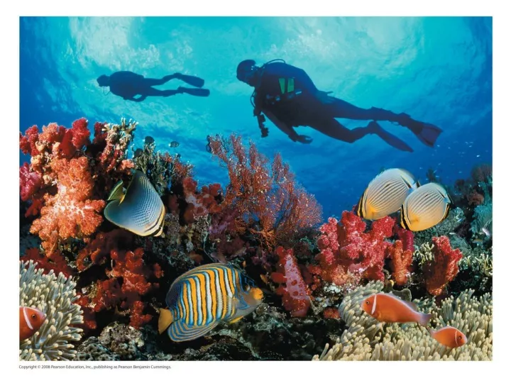
Reproduction and Development • Most animals reproduce sexually, with the diploid stage usually dominating the life cycle • After a sperm fertilizes an egg, the zygote undergoes rapid cell division called cleavage • Cleavage leads to formation of a blastula • The blastula undergoes gastrulation, forming a gastrula with different layers of embryonic tissues Video: Sea Urchin Embryonic Development
Fig. 32-2-1 Cleavage Eight-cell stage Zygote
Fig. 32-2-2 Cleavage Cleavage Blastula Eight-cell stage Zygote Blastocoel Cross section of blastula
Fig. 32-2-3 Blastocoel Endoderm Cleavage Cleavage Blastula Ectoderm Archenteron Eight-cell stage Zygote Gastrulation Gastrula Blastocoel Blastopore Cross section of blastula
Many animals have at least one larval stage • A larva is sexually immature and morphologically distinct from the adult; it eventually undergoes metamorphosis
All animals, and only animals, have Hox genes that regulate the development of body form • Although the Hox family of genes has been highly conserved, it can produce a wide diversity of animal morphology
The history of animals spans more than half a billion years • The animal kingdom includes a great diversity of living species and an even greater diversity of extinct ones • The common ancestor of living animals may have lived between 675 and 875 million years ago • This ancestor may have resembled modern choanoflagellates (single-celled or colonial) that are the closest living relatives of animals
Fig. 32-3 single colonial DNA says “sister groups” Individual choanoflagellate Choanoflagellates OTHER EUKARYOTES Sponges Animals Collar cell (choanocyte) Other animals Identical to collar cells in sponges also found in cniarians, flatworms and echinoderms
Neoproterozoic Era (1 Billion–524 Million Years Ago) • Early members of the animal fossil record date from 565 to 550 million years ago (DNA says older)
Paleozoic Era (542–251 Million Years Ago) • The Cambrian explosion (535 to 525 million years ago) marks the earliest fossil appearance of many major groups of living animals • New predator-prey relationships • A rise in atmospheric oxygen • The evolution of the Hox gene complex
Animal diversity continued to increase through the Paleozoic, but was punctuated by mass extinctions
Mesozoic Era (251–65.5 Million Years Ago) • Coral reefs emerged, becoming important marine ecological niches for other organisms • Dinosaurs! • The first mammals emerged
Cenozoic Era (65.5 Million Years Ago to the Present) • Followed Chicxulub impact!
Probably asteroid, perhaps coupled with vulcanism. Extinctions open niches. Alternative Hypotheses:
Probably asteroid, perhaps coupled with vulcanism. Extinctions open niches.
Concept 32.3: Animals can be characterized by “body plans” • Zoologists sometimes categorize animals according to a body plan, a set of morphological and developmental traits • A grade is a group whose members share key biological features • A grade is not necessarily a clade, or monophyletic group
1. Symmetry • Animals can be categorized according to the symmetry of their bodies, or lack of it • Some animals have radial symmetry
Fig. 32-7 (a) Radial symmetry (b) Bilateral symmetry
Two-sided symmetry is called bilateral symmetry • A dorsal (top) side and a ventral (bottom) side • A right and left side • Anterior (head) and posterior (tail) ends • Cephalization ( to varying degrees), the development of a head
2. Tissues • Tissues are collections of specialized cells isolated from other tissues by membranous layers • During development, 2-3 germ layers give rise to the tissues and organs of the animal embryo
Ectoderm is the germ layer covering the embryo’s surface • Endoderm is the innermost germ layer and lines the developing digestive • Bilateriananimals also have an intervening mesoderm layer
3. Body Cavities • A true body cavity is called a coelom and is derived from mesoderm • Coelomates are animals that possess a true coelom
Fig. 32-8 Coelom Body covering (from ectoderm) Tissue layer lining coelom and suspending internal organs (from mesoderm) Digestive tract (from endoderm) (a) Coelomate Body covering (from ectoderm) Pseudocoelom Muscle layer (from mesoderm) Digestive tract (from endoderm) (b) Pseudocoelomate Body covering (from ectoderm) Tissue- filled region (from mesoderm) Wall of digestive cavity (from endoderm) (c) Acoelomate
Fig. 32-8a Coelom Body covering (from ectoderm) Tissue layer lining coelom and suspending internal organs (from mesoderm) Digestive tract (from endoderm) (a) Coelomate
Fig. 32-8b Body covering (from ectoderm) Pseudocoelom Muscle layer (from mesoderm) Digestive tract (from endoderm) (b) Pseudocoelomate
Fig. 32-8c Body covering (from ectoderm) Tissue- filled region (from mesoderm) Wall of digestive cavity (from endoderm) (c) Acoelomate
4. Protostome and Deuterostome Development • Based on early development, many animals can be categorized as having protostome development or deuterostome development
Cleavage • In protostome development, cleavage is spiral and determinate • In deuterostome development, cleavage is radial and indeterminate
Cleavage • In protostome development, cleavage is spiral and determinate • In deuterostome development, cleavage is radial and indeterminate • With indeterminate cleavage, each cell retains ability to form embryo = identical twins
Fig. 32-9a Deuterostome development (examples: echinoderms, chordates) Protostome development (examples: molluscs, annelids) (a) Cleavage Eight-cell stage Eight-cell stage Spiral and determinate Radial and indeterminate
Fig. 32-9b Protostome development (examples: molluscs, annelids) Deuterostome development (examples: echinoderms, chordates) (b) Coelom formation Coelom Key Ectoderm Archenteron Mesoderm Endoderm Coelom Blastopore Mesoderm Blastopore Mesoderm
Fate of the Blastopore • In protostome development, the blastopore becomes the mouth • In deuterostome development, the blastopore becomes the anus
Fig. 32-9c Protostome development (examples: molluscs, annelids) Deuterostome development (examples: echinoderms, chordates) (c) Fate of the blastopore Anus Mouth Key Ectoderm Digestive tube Mesoderm Endoderm Anus Mouth Mouth develops from blastopore. Anus develops from blastopore.
One hypothesis of animal phylogeny is based mainly on molecular data
Fig. 32-11 Silicea “Porifera” Calcarea ANCESTRAL COLONIAL FLAGELLATE Metazoa Ctenophora Cnidaria Eumetazoa Acoela Echinodermata tissues Deuterostomia Chordata Bilateria Platyhelminthes Rotifera bilateral Ectoprocta Lophotrochozoa Brachiopoda Mollusca protostome Annelida Nematoda Ecdysozoa Arthropoda
Progress in Resolving Bilaterian Relationships • Recent molecular studies indicate three bilaterian clades: Deuterostomia, Ecdysozoa, and Lophotrochozoa • Ecdysozoans shed their exoskeletons through a process called ecdysis
Some lophotrochozoans have a feeding structure called a lophophore • Other phyla go through a distinct developmental stage called the trochophore larva