Download
1 / 54
540 likes | 564 Views
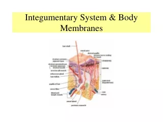
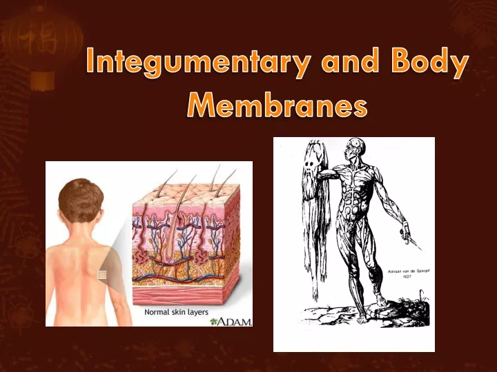
Integumentary and Body Membranes. Body Membranes. Membranes found all throughout body Functions of body membranes: Line or cover body surfaces – separate in/out Protect body surfaces Lubricate body surfaces. Classified into two types: Epithelial membranes Cutaneous membrane

E N D
Body Membranes • Membranes found all throughout body • Functions of body membranes: • Line or cover body surfaces – separate in/out • Protect body surfaces • Lubricate body surfaces
Classified into two types: • Epithelial membranes • Cutaneous membrane • Mucous membrane • Serous membrane • Connective tissue membranes • Synovial membrane
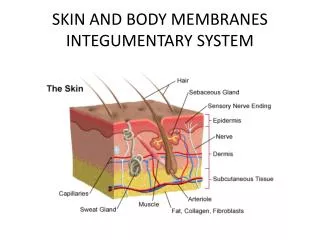
Epithelial Membranes • Cutaneous membrane = skin • Function: protect deeper body tissues • dry membrane • Outermost protective boundary consisting of: • Superficial epidermis • Contains waterproof keratin in areas of high friction • Underlying dermis • Mostly dense connective tissue - protection Figure 4.1a
Mucous membranes • Function: protect from drying out, lubrication • Lines all body cavities that open to the exterior body surface • Often adapted for absorption (i.e. large intestine) or secretion (i.e. nasal cavity)
Serous membranes • Function: lubrication for cushion, friction • Surface simple squamous epithelium • Underlying areolar connective tissue • Lines open body cavities that are closed to the exterior of the body • Serous layers separated by serous fluid
Specific serous membranes • Peritoneum • Abdominal cavity • Pleura • Around the lungs • Inside = visceral • Outside = parietal
Pericardium • Around the heart • Inside = visceral • Outside = parietal
Connective Tissue Membrane • Synovial membrane • Function: lubrication for friction • Connective tissue only • Lines fibrous capsules surrounding joints Figure 4.2
Integumentary System • Four major functions • 1. Protects deeper tissues from: • Mechanical damage • Chemical damage • Bacterial damage • Thermal damage • Ultraviolet radiation • Desiccation (drying out)
2. Aids in heat regulation • Maintain 98.6°F body temperature • 3. Aids in excretion of urea and uric acid • Both nitrogen-based toxins • 4. Synthesizes vitamin D • Needed to help body absorb calcium for bones
Comprised of two things: • Skin (cutaneous membrane) • Skin appendages/derivatives (objects coming from skin) • Sweat glands (sudoriferous glands) • Oil glands (sebaceous glands) • Hairs • Nails
Skin Structure • Epidermis – outer layer • Stratified squamous epithelium (many flat layers) • Often keratinized (hardened by keratin) • Dermis • Dense connective tissue Figure 4.3
Under dermis is the hypodermis • Not part of the skin • Anchors skin to underlying organs • Composed mostly of adipose (connective) tissue • Where blood vessels are located
Layers of Epidermis • Stratum corneum • Shingle-like dead cells; 25-30 layers • Stratum lucidum • Occurs only in thickened skin (calluses, corns) • Stratum granulosum • Stratum spinosum • Stratum basale • Cells undergoing mitosis • Lies next to dermis
Pigment melanin produced by melanocytes is present in epidermis • Color varies from yellows to browns to blacks • Melanocytes are mostly in the stratum basale • Amount of melanin produced depends upon genetics and exposure to sunlight
Layers of Dermis • Papillary layer • Finger-like projections just under epidermis called dermal papillae (form fingerprints) • Pain receptors (nociceptors) at ends of nerves • Capillary beds/loops (blood vessels) where veins & arteries meet • Reticular layer • Blood vessels • Glands • Sensory receptors (Pacinian corpuscle [pressure] and Meissner’s corpuscle [light touch])
Skin Structure Figure 4.4
Skin Color • Three things actually determine skin color: • Melanin • Yellow, brown or black pigments • Carotene • Orange-yellow pigment found in some vegetables that deposits itself in our skin • Eating too many carrots WILL turn skin orange
Susan Dey as Laurie Partridge • During the filming of the show, teenager Susan developed anorexia. • She only ate carrots for weeks at a time. • Eventually, directors had to stop filming because her skin was orange. Don’t Write
Hemoglobin • Red coloring from blood cells in dermis capillaries • Oxygen content determines the extent of red coloring • Light red = oxygenated (arteries) • Dark red = deoxygenated (veins)
Emotional stimuli and/or disease may cause alterations in skin color: • Erythema: redness due to blood vessel dilation • Blushing, hypertension, inflammation, allergy • Pallor: blanching (loss of color) of skin • Emotional stress, low blood pressure, low hemoglobin
Jaundice: yellowing • Excess bile due to liver disorder • Hematomas: bruises
Appendages of the Skin • Many appendages/derivatives of skin. Four major ones: • 1. Sebaceous glands • 2. Sweat (sudoriferous) glands • 3. hair • 4. nails
1. Sebaceous glands • Produce oil called sebum • Lubricant for skin • Kills bacteria • Most with ducts that empty into hair follicles • Glands are activated at puberty
2. Sweat glands • Widely distributed in skin • Two types • Eccrine (merocrine) • Open via duct to pore on skin surface • Apocrine • Ducts empty into hair follicles
Composition of sweat: • Mostly water • Some metabolic waste (i.e. garlic) • Fatty acids and proteins (apocrine only) • Function of sweat: • Helps dissipate excess heat as evaporation occurs • Excretes waste products • Acidic nature inhibits bacteria growth • Odor is from associated bacteria
3. Hair • Produced by hair bulb • Nourished at papilla due to blood vessels • Consists of hard keratinized epithelial cells • Melanocytes provide pigment for hair color Figure 4.7c
Anatomy: • Cuticle • Outermost section • thin, colorless, protection for cortex • Cortex • Middle segment – 90% of hair shaft • strength, color, texture • Central medulla • Innermost segment • only present with thick hairs • Most heavily keratinized structure of body Figure 4.7b
Structures associated with hair: • Hair follicle • Dermal and epidermal sheath surround hair root • Arrector pilli • Tiny smooth muscle causes hair to stand up • Sebaceous gland • Sweat gland • Apocrine only Figure 4.7a
4. Nails • Scale-like modifications of the epidermis • Heavily keratinized • Stratum basale extends beneath the nail bed in matrix • Responsible for growth • Lack of pigment makes them colorless Lee Redmond Guinness World Record Holder – until February 2009 when a car accident broke her nails
Nail structure: • Free edge – what we cut • Body – main nail lying on bed • Root of nail • Eponychium – proximal nail fold that projects onto the nail body (cuticle) • Matrix – stratum basale Figure 4.9
Skin Homeostatic Imbalances • Infections or allergies • Athletes foot • Caused by fungal infection • Boils and carbuncles • Caused by bacterial infection of hair follicle • Cold sores • Caused by viral infection • Herpes simplex I
Contact dermatitis • Exposures cause allergic reaction • Eczema (atopic dermatitis) • Hypersensitivity reaction (allergy) • Most common in infants & many outgrow it • Impetigo • Caused by bacterial infection usually a result of eczema • Psoriasis • Cause is unknown • Triggered by trauma, infection, stress
Onycholysis • Separation of nail from nail bed • Symptom of trauma or infection • Cyanosis • Bluish (cyan) coloring • Symptom of inadequate oxygen in the blood • Common in newborns • Vitiligo • Loss of pigmentation • Melanocytes destroyed
Pressure ulcers (bedsore) • Lack of unrelieved pressure, friction, humidity, temperature, age, continence and medication • Disrupts blood flow & oxygen to cells, killing them Decubitus ulcers
Burns • Tissue damage and cell death caused by heat, electricity, UV radiation, or chemicals • Associated dangers • Infection • Dehydration • Electrolyte imbalance • Circulatory shock
Way to immediately determine the extent of burns is with the “Rule of Nines” • Body is divided into 11 areas for quick estimation • Each area represents about 9% • One side of leg = 9% • One whole arm = 9% Figure 4.11a
Burns are categorized by severity in degrees • First-degree burns • Only epidermis is damaged • Skin is red and swollen • Second-degree burns • Epidermis and upper dermis are damaged • Skin is red with blisters • Third-degree burns • Destroys entire skin layer • Burn is gray-white or black Epidermis Dermis 3° 2° 1°
Burns are considered critical if: • Over 25% of body has second degree burns • Over 10% of body has third degree burns • Third degree burns on face, hands, or feet
Skin cancer – abnormal cell mass (tumor) of epidermis • Two types of tumors • Benign • Does not spread (encapsulated) • Slow-growing • Malignant • Can metastasize (moves) to other parts of the body • Fast-growing (starve other cells) Epidermis Dermis
Three types of skin cancer: • Basal cell carcinoma • Least malignant • Most common type • Arises from stratum basale • Squamous cell carcinoma • Arises from stratum spinosum • Metastasizes to lymph nodes • Early removal allows a good chance of cure
Malignant melanoma • Cancer of melanocytes • Most deadly of skin cancers • Metastasizes rapidly to lymph and blood vessels
Detection of skin cancer uses ABCDE rule • A = Asymmetry • Two sides of pigmented mole do not match • B = Border irregularity • Borders of mole are not smooth • C = Color • Different colors in pigmented area • D = Diameter • Spot is larger than 6 mm in diameter • E = Evolving • Changing over time in size, color, texture
Chemotherapy affects integumentary system more than any other system • Drugs target rapidly dividing cells (cancer) • Integument has rapidly dividing cells (stratum basale) in matrix area • Loss of hair – destroys matrix • Dry skin – destroys stratum basale • Dry brittle nails – destroys matrix • Dry mouth & throat – destroys mucus membranes • Nausea – destroys mucus membranes in stomach
Development • Many changes in integumentary system occur from conception to death • Hair changes • Skin changes