Download
1 / 54
540 likes | 576 Views
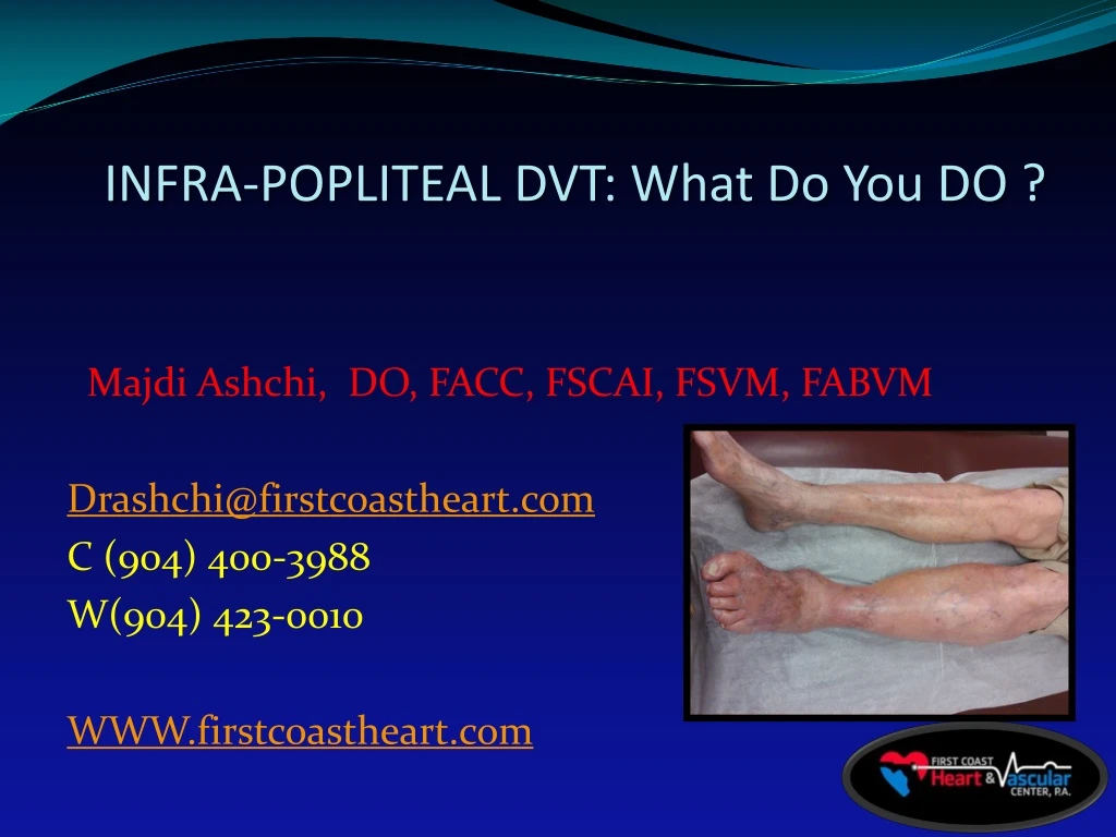
INFRA-POPLITEAL DVT: What Do You DO ?. Majdi Ashchi, DO, FACC, FSCAI, FSVM, FABVM Drashchi@firstcoastheart.com C (904) 400-3988 W(904) 423-0010 WWW.firstcoastheart.com. DISCLOSURES. NONE. OBJECTIVES:. Brief Discussion : VTE

E N D
INFRA-POPLITEAL DVT: What Do You DO ? Majdi Ashchi, DO, FACC, FSCAI, FSVM, FABVM Drashchi@firstcoastheart.com C (904) 400-3988 W(904) 423-0010 WWW.firstcoastheart.com
DISCLOSURES • NONE
OBJECTIVES: Brief Discussion : • VTE • Discuss Briefly The Controversy & Literature And Guidelines For The DX, Treatment of Proximal DVT vs Distal DVT ( Infra-Popliteal) • Diagnostics Modalities of DVT • Epidemiology of VTE • Risk Factors For VTE
https://www.facebook.com/majdi.ashchi/posts/1674122196142090
VTE- Venous Thromboembolism • VTE –pathologic thrombosis that occurs within the venous circulation. • Most common form of VTE is DEEP VENOUS THROMBOSIS (DVT) of the lower extremity • Most Life threatening manifestation is embolization of thrombus resulting in Pulmonary Embolism. • VTE is often SILENT disease. • Accurate assessment of clinical symptoms, signs, risk factors, and appropriate use of objective diagnostic tests is important in the accurate diagnosis, prevention and treatment of VTE.
Epidemiology of VTE • DVT is a major USA clinical problem • Incident 500k-2 million cases per year • ICD-9 for DVT showed 250K admissions per year • Estimated LOS 24 hours to 7 days. • Major 3 complications: • Recurrent non-fatal VTE • Post-thrombotic Syndrome • Fatal Pulmonary Embolism
Epidemiology VTE • GOAL of therapy of DVT is Prevention Of: • Thrombus propagation • Thrombus embolization • Early & Late thrombus recurrence • Proper Anticoagulation is the first critical step in effective treatment of DVT. • Complications can develop soon after DVT detection which gives a narrow window of opportunity for safe and effective intervention • Secondary stage of treatment involves maintenance of sufficient anticoagulation to prevent recurrence of DVT
ETIOLOGY- Risk Factors https://www.youtube.com/watch?v=LvfPgKgsi-w Virchow’s Triad characterizes the development of thrombosis as a result of a combination of venous stasis , venous endothelial injury & hypercoagulability. Understanding risks for developing VTE can help determine choice of prevention modalities, as well as diagnosing of VTE when patients present with signs & symptoms of thromboembolic disease. Risk factors can be both acquired or inherited, & combinations of risk factors are often present. • Common risk factors: • History of VTE • Immobility • Hospitalization • Surgery • Malginancy • Infection • Thrombophilia
Virchow’s Triad Burns Fractures ,Inherited or aquired
RISK FACTORS • Immobility • Major Surgery (THR, TKR, Hip Fx, CA Sx, Abdominal operations requiring general anesthesia > 30 min) • Trauma • Lower Extremity Paresis • Sedentary Life Style • Long Distance Travel • Medical Illness • CHF OSA Cancer Liver Disease • Nephrotic Syndrome • Previous Venous Thrombosis • (x 8 fold if hospitalized) • Obesity • Autoimmune & Inflammatory Disorder • Hematologic & Myloproliferative Disorder • Polycythemia Vera • Paroxysmal Nocturnal Hemoglobinuria • Inherited Or Acquired Thrombophilia • Factor V leiden mutation • Prothrombin gene Mutation • Protein S & C Deficiency • Antithrombin III Deficiency • HIT • Aniphospholipid Antibody Syndrome • External Compression Syndromes • May-Thurner Syndrome • Paget-Schroetter Syndrome • Hematoma • Tumor • Endovascular Devices • Central Venous catheters • Hemodialysis Catheters • IVCF (risk for DVT_ • Pacemaker/Defibrillator Wires • Extracorporeal membrane Oxygenation catheters • Demographic/Behavioral • Age • Smoking • Sedentary Lifestyle • Long distance travel • Pregnancy • Drug Induced • Chemotherapy • Estrogen Containing Oral Contraceptives or HRT • Selective Estrogen Modulators (eg. Tamoxifen)
Signs & Symptoms of DVT • Majority Of Patients with DVT are ASYMPTOMATIC • Edema • Erythema • Tenderness with dorsiflexion of foot (Homan’s sign) • Calf pain on palpation ( Pratt Sign) • Both signs have S&S <50% • Bluish Discoloration
Diagnosis Of DVT (1) • Traditional Clinical Criteria to diagnose PE and DVT for the lower extremity are neither S nor S and have poor positive predictive value. • Highest diagnostic accuracy achieved when pretest clinical assessment tools (i.e. Wells criteria) are combined with objective biomarker and image testing in defined management strategies.
Diagnosis Of DVT (2) Wells Criteria ( point system) • Combining the Wells clinical predicted model with a single duplex us (DUS) is a management strategy that yields a significant improvement on the diagnostic accuracy: Positive pretest probability of venous thromboembolism of • 3% in Low risk • 16% in Moderate risk • 74.6% in High risk • Patients with Negative US for DVT, frequency of thromboembolic events in 3 months is only 0.6%.
D-Dimer • Fibrin degradation product elevated in active thrombosis • ELISA-Enzyme linked immunosorbent assay-most available & validated. • Clinical value of D-dimer relies on its high negative predictive value. • D-Dimer level <0.5 ng/dl or less in the setting of a low or intermediate pretest probability (eg Wells score <=2) confers a risk of PE or DVT of less than 1% in 3 months. • Positive D-Dimer with a high pretest probability for PE or DVT, should prompt the clinician to proceed with imaging.
Duplex Ultrasound • Reliable method for evaluating symptomatic proximal DVT. It is considered diagnostic for proximal DVT. • Several criteria but Non compressibility is the most accurate diagnostic feature with >95% S&S when compared to venography. • DUS is less reliable in diagnosis of isolated calf DVT, with S & S <70%.
Contrast Venography • Reference standard • Invasive, so less favored • Useful in areas where DUS shown poor diagnostic accuracy (IVC, Iliac Vein, Calf veins) • Can cause nephropathy, Induced superficial thrombophlebitis, DVT, and can be used with dye limitation and limb limitation.
CTV -computed Tomography Venography • Used or preferred when DUS is non-diagnostic • Similar to MRV in showing anatomy veins etc. • NOT first line role diagnostic study for DVT • Significant External Beam Radiation • Preferred for lungs in PE ( superior to V/Q scan) • Contra-indicated in renal failure, DM, CHF
MRV-Magnetic Resonance Venography • Comparable to angiography in S&S • Used where DUS is not preferred or less sensitive • MRV allow simultaneous leg imaging • Provides accurate venous flow information i.e. Iliac veins, IVC ( central venous anatomy) • Aids to differentiate Acute from remote DVT. • 10% of patients can not have it due to metals in body or claustrophobia.
DVT : proximal vs Distal • PROXIMAL: located in the popliteal, femoral, or iliac veins. • DISTAL : Isolated distal DVT encompasses those located below the knee in the calf veins . ( popliteal vein is not involved). Most calf DVT are located in the posterior Tibial and peroneal veins while AT vein and muscular vein DVTs are uncommon.
INFRAPOPLITEAL DVT-(distal or calf) DOES IT EVEN MATTER AND SHOULD WE TREAT IT ???
Infra-Popliteal DVT Is anticoagulation necessary to preventa pulmonary embolus ?
GUIDELINES : • Isolated calf DVT can be managed only with serial duplex US surveillance ? • TRUE • FALSE • None of the above
GUIDELINES of Infrapoplitealtx • Isolated calf DVT are treated with anticoagulation only • TRUE • FALSE • None of the above
Guidelines Surveillance US Anticoagulation • NO risk factors • Without severe symptoms • Risk factors for extension • Severe symptoms
Anticoagulation VS Surveillance:why each? Anticoagulation Surveillance • Reduce risk of propagation/embolization • No Need for Multiple return visits for DUS • ? Reduction in long term symptoms of venous insufficiency • NO risk of anticoagulant induced hemorrhage • Able to keep close tabs on the patient/symptoms • Risk of propagation/embolization is low.
PROXIMAL DVT LANCET 1960; 1: 1309 Seminal Trial Ann Intern Med 2010;152:578 • Anticoagulation Vs Observation of acute DVT resulted in dramatic reduction in recurrence and mortality benefit. • Meta-analysis of 13 prospective cohort studies and 56 randomized clinical trials reported recurrent VTE 3.4% and fatal VTE 0.4% during first three months of anticoagulant therapy.
Propagation of DVT during Surveillance NO prophylaxis J V Surg 2012;55;550-61
Infra-Popliteal DVT-what do we know? DVT is a common problem among hospitalized & Recently discharged patients. ( we all agree to this) Proximal DVT requires therapy due to prohibitive risk of propagation with just surveillance. Indication to anticoagulate for proximal DVT is stronger than distal because risk of complications is higher, especially embolization and death. 90% of acute PE arise from proximal veins. OPTIMEDV Study showed mortality rate for proximal DVT is higher than for distal DVT (8 %vs 4 %). Lancet 1974; 1:258 ActaChirScandSupp 1977;478: 1 ThrombHaemost 2009; 102: 493
Distal or Infrapopliteal DVT • What about calf DVT? Why Should we even care? • Many Vascular Laboratories or hospitals do NOT perform tibial vein duplex ultrasound as part of the imaging protocol. It has low S&S. • Literature here is NOT as clear cut • We know from guideline, we have symptomatic & asymptomatic patients with distal DVT to deal with.
DVT: Surveillance with DUS • The optimal frequency, duration and method of surveillance are unknown • Patients may have surveillance: • High risk for bleeding or those with preference • Support for surveillance comes from studies that suggest that the risk for embolization of isolated distal DVT is low and approximately half of that of proximal DVT. • BMJ :2011; 342: d3036
DVT: Surveillance with DUS 1( support for surveillance) • Prospective observational and retrospective studies reported that limited thrombosis of muscular veins compared with extensive thrombosis of multiple calf veins has low risk of extension without therapy (3 vs 15%). Also, if extension does not occur within two weeks, it is unlikely to occur. • J VascSurg 2010;52 :1246 & 1251 • J Vasc Sur 2003; 37: 523
6 Randomized Controlled Trials for Calf DVT J V Surg 2012;55;550-61
Meta-analysis 2 R & 6 non-R trials • Patients with isolated distal DVT • Compared those who were followed with serial DUS • Anticoagulated patients were less likely to have proximal DVT propagation ( odd ratio 0.29, 95% CI). • However, methodologic quality of most studies was poor and the number of outcome events that occurred (death, PE, proximal DVT extension, bleeding) was small, which limited the analysis. J V Surgery 2012; 55:550-561
Distal DVT-indications to anticoagulation • Natural History suggest that when left untreated, 1/3 with symptomatic isolated distal DVT will develop extension into proximal veins, most often within first two weeks after diagnosis. • Circulation 2003;107:122 • J VascSurg 2003;37:523 • Support for anticoagulation is based on risk of extension into proximal veins where anticoagulation has stronger indication due to higher risk for embolization and proven efficacy in this population in reducing clot extension.
Distal DVT- Indication for anticoagulation (both 1) Asymptomatic distal DVT Patient who become symptomatic with distal DVT who opted for surveillance • DVT extension into or toward the proximal vein during surveillance • Patient considered by clinician to be at risk for extension to the proximal veins which includes: • Unprovoked DVT • D-Dimer >500 mg/ml • Extensive thrombosis involving multiple veins (>5cm length and >7mm in diameter) • Thrombosis close to the proximal veins. • (Continue) • Inpatient status • Prolonged immobility • Prior DVT or Pulmonary E • Peristent/Irreversible Risk factors such as active cancer. • Chest 2012; 141: e419S.
Symptomatic Distal DVT Most clinicians agree that anticoagulation is indicated in isolated DVT provided risk of bleeding is low. Chest 2012; 141: e419S. Blood 2014; 124: 196 Chest 2014; 146: 1468
Etiology (risk assessment 3) • ACCP 2008 Guidelines for prevention of VTE advocate for a group specific approach to VTE risk assessment by assigning risk according to the type of surgery, mobility, & individual risk factors ( see table of risk factors). • Chest. 2008; 133 (6 suppl): S381-S453. • Caprini Risk Assessment Model • Validated via general & urological surgical populations • SeminThrombHemost 1991;17(suppl 3):304-312 • Rogers Score For Surgical Patients. • Cumbersome risk assessment tool & has not been validated. • J Am Coll Surg. 2007;204(6):1211-1221
Risk Stratification for VTE • LOW risk • Mobile patient, having minor surgery • Medical patient who is fully ambulatory • <10% without thrombophylaxis • MODERATE risk • Patients undergoing general, open gynecologic or urologic surgery • Medically Ill patients with =>2 risk factors • 10-40% risk of VTE without thrombophylaxis. • HIGH risk • Patient with hip, knee replacement, fractured hip surgery • Major Trauma, acute spinal cord injury, major cancer surgery • 40-80% risk for VTE without thrombophylaxis