Download
1 / 53
610 likes | 1.16k Views
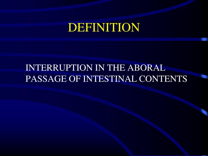
DEFINITION. INTERRUPTION IN THE ABORAL PASSAGE OF INTESTINAL CONTENTS. The common Scenario. A 50 year old man presents with abdominal pain, distension, obstipation and constipation, with repeated episodes of vomiting.

E N D
DEFINITION INTERRUPTION IN THE ABORAL PASSAGE OF INTESTINAL CONTENTS
The common Scenario A 50 year old man presents with abdominal pain, distension, obstipation and constipation, with repeated episodes of vomiting. His vital sign were stable, abdomen distended with diffuse tenderness. Bowel Sounds are hyperactive.
Clinical Picture • Colicky abdominal pain • Abdominal distension • Vomiting • Decreased passage of stool or flatus • Typical radiographic picture • plain AXR, contrast CT, UGI/SBFT, enteroclysis
Gas diffusely through intestine, incl. colon May have large diffuse Air/fluid levels Quiet abdomen No obvious transition point on contrast study Distended small bowel loops, no air in colon Laddered Air/fluid levels Active peristalsis, quiet= late Obvious transition point on contrast study Paralytic vs Mechanical Ileus Obstruction
Upright Abdominal Film Air - Fluid Levels Dilated Small Bowel
Pathophysiology I • Hypercontractility--hypocontractility • Massive fluid losses: • Vomiting • sequestration of fluid into the lumen from the surrounding circulation • Lymphatic and venous congestion resulting in oedematous tissues - oliguria, hypotension, hemoconcentration • Electrolyte depletion
Pathophysiology II • bowel distension--increased intraluminal pressure--impediment of venous return--arterial insufficiency--bowel wall ischemia --necrosis- -perforation--peritonitis and sepsis • Bacterial over growth with translocation of bacteria and it’s toxins causing bacteraemia and septicaemia.
Important Questions • Site • Etiology • Partial vs. complete • Simple vs. strangulated • Fluid & electrolyte status • Operative vs. non-operative management
Site – Small Bowel May occur at any point in length of small bowel
Site? Small Bowel vs. Large Bowel • Scenario • prior operations, in bowel habits • Clinical picture • scars, masses/ hernias, amount of distension/ vomiting • Radiological studies • gas in colon?, volvulus?, transition point, mass • (Almost) always operate on LBO, often treat SBO non-operatively
Etiology? • Outside the wall • Inside the wall • Inside the lumen
Small Bowel Adhesions • Account for 60-70% of All SBO • Result from peritoneal injury, platelet activation and fibrin formation. • Associated with starch covered gloves, intraperitoneal sepsis, haemorrhage and foreign bodies. • As early as 4 weeks post laparotomy. The majority of patients present between 1-5 years • Colorectal Surgery 25% • Gynaecological 20% • Appendectomy 14% • 70% of patients have a single band • Patients with complex bands are more likely to be readmitted • Readmission in surgically treated patients is 35%
Hernia • Accounts for 10% of SBO • Commonest 1. Femoral hernia 2. Inguinal 3. Umbilical 4. Others: Incisional and Internal H. • The site of obstruction is the neck of the hernia • The compromised viscus is within the sac. • Ischaemia occurs initially by venous occlusion, followed by oedema and arterial compromise. • Strangulation is noted by: • Persistent pain • Discoloration • Tenderness • Systemic symptoms
Other causes Intussusception Gall stone Ileus IBD
frequency Common Causes of LBO • Colon cancer • Diverticulitis • Volvulus • Hernia Unlike SBO, adhesions very unlikely to produce LBO
Sigmoid Volvulus Colonic Obstruction
Flatus Residual colonic gas above peritoneal reflection Adhesions 60-80% resolve with non-operative Tx Must show objective improvement, if none by 48h consider OR Complete obstipation No residual colonic gas on AXR SBFT may differentiate early complete from high-grade partial Almost all should be operated on within 24h Partial vs Complete
Is there strangulation? • 4 Cardinal Signs fever, tachycardia, localized abdominal tenderness, leukocytosis • 0/4 0% strangulated bowel • 1/4 7% “ “ • 2-3/4 24% “ “ • 4/4 67% “ “ • process accelerated with closed-loop obstr.
Role of CT • Used with IV contrast, oral and rectal contrast (triple contrast). • Able to demonstrate abnormality in the bowel wall, mesentery, mesenteric vessels and peritoneum. • It can define • Level of obstruction • Degree of obstruction • Cause: volvulus, hernia, luminal and mural causes • The degree of ischaemia • Free fluid and gas • Ensure: patient vitally stable with no renal failure and no previous allergy to iodine
Pneumatosis Intestinalis • Dilated Loops of SB • Air in Wall of SB • No Air in Colon
Role of barium& gastrografin studies Barium should not be used in a patient with peritonitis • As: UGI with follow through, enema • Gastrografin is used in acute abdomen but is diluted • Useful in recurrent and chronic obstruction • May be able to define the level and mural causes. • Can be used to distinguish between adynamic and mechanical obstruction
Causes of Adynamic Ileus • Following celiotomy • small bowel- 24h, stomach- 48h, colon- 3-5d • Inflammation e.g. appendicitis, pancreatitis • Retroperitoneal disorders e.g. ureter, spine, blood • Thoracic conditions e.g. pneumonia, # ribs • Systemic disorders e.g. sepsis, hyponatremia, hypokalemia, hypomagnesemia • Drugs e.g opiates, Ca-channel blockers, psychotropics
Management of Bowel Obstruction NEVER LET THE SUN RISE OR FALL ON A PATIENT WITH BOWEL OBSTRUCTION
Principles • Fluid resuscitation • Electrolyte, acid-base correction • Close monitoring • foley, central line • NGT decompression • Antibiotics controversial • TO OPERATE OR NOT TO OPERATE
Resuscitation • Massive third space losses as fluid and electrolytes accumulate in bowel wall and lumen • Depending on site and duration • Proximal - vomiting early, with dehydration, hypochloremia, alkalosis • distal- more distension, vomiting late, dehydration profound, fewer electrolyte abnormalities • Requirements = DEFICIT + MAINTENANCE + ONGOING LOSSES
When is it safe NOT to operate? • SMALL bowel obstruction if adhesions suspected etiology i.e. CANNOT have a “virgin” abdomen • No signs of strangulation • Adynamic ileus
Operative Indications • Incarcerated or strangulated hernia • Peritonitis • Pneumopertioneum • Suspected strangulation • Closed loop obstruction • Complete obstruction • Virgin abdomen • LARGE bowel obstruction
Case 1 • 82yo man /c CHF and Hairy Cell Leukemia. Presents to the ER /c dx of appendicitis. Taken to the OR for uncomplicated laparoscopic appendectomy. • POD #2 - progressive abdominal distention with postop ileus • POD#3 - bilious emesis - afeb, nontender abd, wcc 5000
Case 1 • POD#5 - Abdomen distended - High NGT output - No classic signs of strangulation
Outcome 1 • Taken to OR for laparoscopic exploration evening of POD#5 • Findings: • Suture at umbilical Hasson trocar site had broken (knot intact) • Richters hernia • Proximal bowel viable but congested • Peristalsis, doppler signal and Wood’s lamp all negative for ischemic injury
Case 2 • 60yo M s/p R hemicolectomy 9/99 for cancer. Presents with 3d of intermittent crampy epigastric pain, distension, n/v. 3 “normal” BMs in 24 hours. • PE: T36.8 141/91 92 18 • Absent BS, soft, distended abdomen with periumbilical tenderness. No rebound or guarding. No palpable hernias. Well healed scars. • Labs: WBC 15.7, Hct 48, HCO3 28 nl LFTs and amylase Negative UA
Outcome 2 • NGT placed, fluid resuscitated. • Given high grade obstruction on AXRs, and leukocytosis patient taken to OR within 24 hours. • On laparotomy, multiple dense adhesions found with tight band in retroperitoneum causing internal hernia/obstruction with a transition point. Adhesiolysis performed.
Case 3 • 79yo F with Parkinson’s disease and h/o breast cancer 20 yr ago presents to with 4d h/o n/v, distension. No abd pain. Reports recent bowel movement • PE: Afebrile BP157/74 P89 Hard palpable mass in RUQ. Distended abdomen, high pitched BS, no tenderness. No palpable hernias. No scars. Black stool. • Labs: WBC 10.1 Hct 23.8 Cr 0.7 LFT’s wnl
Outcome 3 • Operative exploration given RUQ mass, abd CT demonstrating distended small bowel and decompressed colon, with multiple masses in the RUQ and pelvis. • On laparotomy, large RUQ mass involving multiple loops of small and large bowel, and mass in R pelvis requiring small and large bowel partial resections. Pathology lobular adenocarcinoma. Regained bowel function POD 5.
Case 4 • 32 ym, former athlete • Ex lap for ruptured appendix 1997 • Non-operative management partial SBO 2002 • Presents to ER four months later w/ diffuse abdominal pain and distension • PE: T 36.5, HR 75, mild periumbilical tenderness, no peritonism, midline scar, reducible LIH • Labs: WCC 13.5, HCO3 25, other labs WNL