Download
1 / 1
10 likes | 119 Views
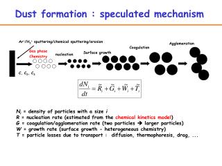
Molecular Mechanism of Hydrogen-Formation in Fe-Only Hydrogenases. Nicolai Lehnert, Department of Chemistry, University of Michigan, Ann Arbor, MI 48109. Hydrogenase models: [Fe 2 (edt)(PMe 3 ) 4 (CO) 2 (H)] + terminal and bridging hydride isomers.

E N D
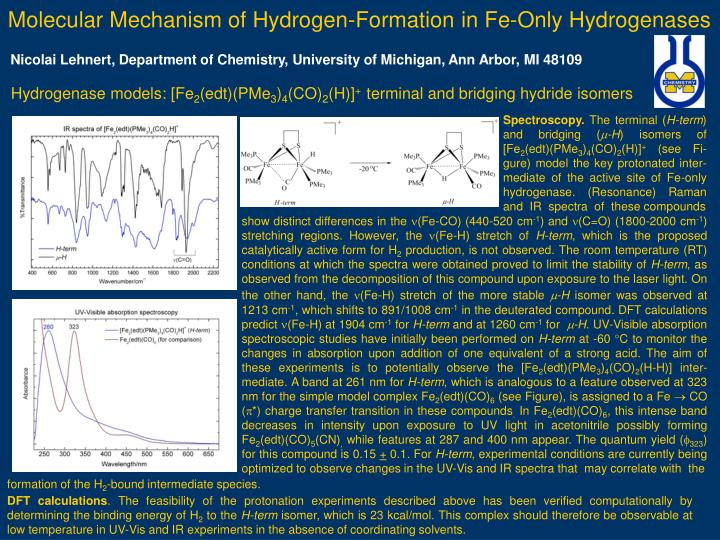
Molecular Mechanism of Hydrogen-Formation in Fe-Only Hydrogenases Nicolai Lehnert, Department of Chemistry, University of Michigan, Ann Arbor, MI 48109 Hydrogenase models: [Fe2(edt)(PMe3)4(CO)2(H)]+ terminal and bridging hydride isomers Spectroscopy. The terminal (H-term) and bridging (m-H) isomers of [Fe2(edt)(PMe3)4(CO)2(H)]+ (see Fi-gure) model the key protonated inter-mediate of the active site of Fe-only hydrogenase. (Resonance) Raman and IR spectra of these compounds show distinct differences in the n(Fe-CO) (440-520 cm-1) and n(C=O) (1800-2000 cm-1) stretchingregions. However, the n(Fe-H) stretch of H-term, which is the proposed catalytically active form for H2 production, is not observed. The room temperature (RT) conditions at which the spectra were obtained proved to limit the stability of H-term, as observed from the decomposition of this compound upon exposure to the laser light. On the other hand, the (Fe-H) stretchofthe more stable -H isomer was observed at 1213 cm-1, which shifts to 891/1008 cm-1 in the deuterated compound. DFT calculations predict n(Fe-H) at 1904 cm-1 for H-term and at 1260 cm-1 for m-H. UV-Visible absorption spectroscopic studies have initially been performed on H-term at -60 C to monitor the changes in absorption upon addition of one equivalent of a strong acid. The aim of these experiments is to potentially observe the [Fe2(edt)(PMe3)4(CO)2(H-H)] inter-mediate. A band at 261 nm for H-term, which is analogous to a feature observed at 323 nm for the simple model complex Fe2(edt)(CO)6 (see Figure), is assigned to a Fe CO (*) charge transfer transition in these compounds. In Fe2(edt)(CO)6, this intense band decreases in intensity upon exposure to UV light in acetonitrile possibly forming Fe2(edt)(CO)5(CN), while features at 287 and 400 nm appear. The quantum yield (323) for this compound is 0.15 + 0.1. For H-term, experimental conditions are currently being optimized to observe changes in the UV-Vis and IR spectra that may correlate with the formation of the H2-bound intermediate species. DFT calculations. The feasibility of the protonation experiments described above has been verified computationally by determining the binding energy of H2 to the H-term isomer, which is 23 kcal/mol. This complex should therefore be observable at low temperature in UV-Vis and IR experiments in the absence of coordinating solvents.