Download
1 / 52
540 likes | 803 Views
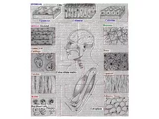
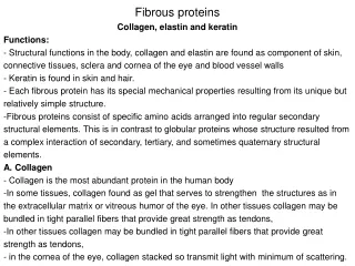
Chapter 8 Fibrous proteins Major fibrous protein of epithelial tissues is a keratin Major fibrous proteins of connective tissue are: Collagen Elastin Dr. Stephen C. Hardies 7-3735 437D hardies@uthscsa.edu.

E N D
Chapter 8 Fibrous proteins Major fibrous protein of epithelial tissues is a keratin Major fibrous proteins of connective tissue are: Collagen Elastin Dr. Stephen C. Hardies 7-3735 437D hardies@uthscsa.edu
The fundamental building block of a keratin fiber is a coiled coil dimer of a type I and type II keratin polypeptide. A. a-helical coiled coil domain B. coiled coil a-helix heptad repeat: hxxhxxx, where "h" means hydrophobic and "x" means any residue. C.
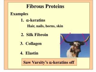
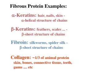
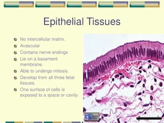
a-Keratin Where: Major protein of hair, skin, nails, some oral mucosa; small amounts in all epithelial cells. Cellular location: Intracellular; not found in connective tissues Structure: fibrous bundle of coil-coiled a-helixes. Function: Provide insoluble structural protein for body surfaces Crosslinks: disulfide bonds Multigene family: family members differ by: tissue structure Hard Soft amt. Cys More Less amt. Crosslinks More Less
True or False? http://biochem.uthscsa.edu/hardies-bin/survey.pl a) Proteins are linear chains of amino acids with polarity. Polarity means that there is directionality caused by adjacent amino acids being joined from COOH group to NH2 group. One end of a polypeptide will be termed the N-terminus and the other the C-terminus. • Disulfide bonds are crosslinks between sulfur atoms of either • cysteine residues or methionine residues. • c) An alpha helix can accommidate any sequence of amino acids.
Collagen Where: 30% of total protein. Major protein of connective tissues: bones, tendons, ligaments, basement membranes, dentin, cementum, (not enamel). Cellular location: extracellular matrix. Structure: triple helix (tropocollagen). Subsequent to secretion, tropocollagen is assembled and crosslinked to make insoluble collagen fibers. Function: Provides tensile strength to soft connective tissues. Tissues that must be elastic but exhibit tensile strength (e.g. ligaments) have a mixture of collagen and elastin. Collagen fibers in bone reinforce against fracture.
True or False? http://biochem.uthscsa.edu/hardies-bin/survey.pl a) In woven bone, collagen fibers are laid down in a disorganized array, whereas in lamellar bone they arelaid down in a more organized parallel fashion. b) Woven bone is stronger than lamellar bone. c) Laminar bone is made by remodeling woven bone.
Features of collagen primary structure Repetitive character of sequence: -Gly-Pro-Met-Gly-Pro-Ser-Gly-Pro-Arg- -Gly-Leu-Hyp-Gly-Pro-Hyp-Gly-Ala-Hyp- -Gly-Pro-Gln-Gly-Phe-Gln-Gly-Pro-Hyp-
The collagen triple helix is stabilized by an interchain hydrogen bonding network involving the hydroxyl group of hydroxyproline, the glycine carbonyl group, and water molecules.
True or False? http://biochem.uthscsa.edu/hardies-bin/survey.pl • Hydroxyproline and hydroxylysine are among the 20 amino acids inserted into polypeptides by ribosomes. • Glycine is NH2-CH2-COOH. • Glycine is the only amino acid that doesn’t have a D and an L isomer.
Type I collagen fibril EM Gap tropocollagen Key assembly interaction 670 angstrom D period
Ca10(OH)2(PO4)6 gap gap gap gap
True or False? http://biochem.uthscsa.edu/hardies-bin/survey.pl • Ca10(OH)2(PO4)6 is the same mineral (hydroxyapatite) that forms enamel. • Whereas bone is reinforced by collagen fibers, enamel uses a different protein named amelogenin. • Enamel has a higher mineral content than bone.
fibril Type VI Anchoring fiber 5 nm 50 nm
Type IV collagen forms planar arrays and makes basement membranes Type VII collagen anchors basement membrane to underlying connective tissue (stromal) cell layer.
Type III collagen Sometimes called elastic collagen or extensible collagen
Collagen type III micrograph visualized in polarized light, showing crimped organization of the fibrils.
Defects in Type I collagen cause: Osteogenesis Imperfecta Dentinogenesis Imperfecta Opalescent and cracked teeth Blue sclera Type I: associated with OI. Will probably need full crown coverage.
An example of a mutation underlying Osteogenesis Imperfecta 988 Pro Gly Pro Arg Gly Arg Thr Gly Asp Ala CCG GGT CCT CGC GGT CGC ACT GGT GAT GCT Pro Cys Pro Arg Gly Arg Thr Gly Asp Ala CCT TGT CCT CGC GGT CGC ACT GGT GAT GCT
True or False? http://biochem.uthscsa.edu/hardies-bin/survey.pl • DNA is a polymer of bases named A,T,G, and C. • The sequence of bases in a gene determines the sequence of the polypeptide that will be produced. • A “mutation” is a heritable change to the sequence of a gene that causes the encoded polypeptide to have altered function. • Some individual residue changes may have large effects on the protein function, whereas other changes may have little or no effect. • Sickle cell anemia is an example of an inherited disease caused by a single residue change in a protein.
Ehlers-Danlos Syndrome Hyperplasticity of the skin
Steps in collagen biosynthesis: • Translation on rough ER and entry into ER. • Hydroxylation in the ER. • Triple helix assembly. • Glycosylation, transport to Golgi, further glycosylation. • Secretion. • Removal of propeptides. • Assembly into fibrils. • Crosslinking.
Scurvy Perifollicular abnormalities. Gingival abnormalities
True or False? http://biochem.uthscsa.edu/hardies-bin/survey.pl What are preliminary steps to get procollagen polypeptides into the ER? • Depending on the synthetic cell type, transcription factors willselect specific preprocollagen genes from a family of such genes be transcribed and translated. • The preprocollagen polypeptide is released into the cytoplasm. • A special sequence on the N-terminus called the ‘signal peptide’directs the preprocollagen to a pore in the ER through which itenters the ER.
Examples of collagen nomenclature: An individual polypeptide:a1(I) procollagen Assembled triple helix with propeptides still on: Type I procollagen Triple helix after propeptide removal: Type I tropocollagen Assembled into 50 nm fiber: Collagen microfibril
A third lysine (from a third tropocollagen) can add to make pyridinoline. Crosslinks involving hydroxylysine are more stable than those involving lysine. There is also a crosslink involving histidine.
Steps in collagen biosynthesis: • Translation on rough ER and entry into ER. • Hydroxylation in the ER. • Triple helix assembly. • Glycosylation, transport to Golgi, further glycosylation. • Secretion. • Removal of propeptides. • Assembly into fibrils. • Crosslinking. vitamin C + HOlys HOpro (or else degrade) + + +
Elastin: Where: elastic connective tissues. Cellular location: extracellular matrix Function: add elasticity to connective tissue Structure: beta spiral Crosslink: desmosine
Genetic defects in elastin underlie Williams Syndrome Facial features associated with Williams Syndrome Dental features include small widely spaced teeth and malocclusion.
Dynamic structure of elastin: The structure of elastin is called a b spiral, and is loosely held together by the hydrophobic force. It is easily deformed to an extended configuration, but will relax back to a compact conformation. Repeating unit: PGVGV
Fibrillin Where: connective tissue; extracellular matrix Structure: forms outer envelope of elastin microfibrils Genetic defects in fibrillin result in Marfan’s Syndrome, characterized by a tall gaunt appearance, joint problems, an often leading to death by aortic aneurysm.
Enzymes that turn over connective tissue Common names: collagenase, gelatinase, elastase Formal names: Matrix Metalloproteases (MMPs) Function: degrade connective tissue in support of tissue remodeling, wound healing, and cell migration (including during metastasis). Family of Zn++ -requiring zymogens embedded in connective tissue. They can be activated in a cascade starting from a cell surface MMP.
True or False? http://biochem.uthscsa.edu/hardies-bin/survey.pl • Zymogens are inactive forms of enzymes that will become activated by cleavage to remove a propeptide. Zymogenes are also called “proenzymes”. Examples of zymogens are: • trypsinogen • collagen • coagulation factors • DNA polymerase • cathepsin
Proteoglycan aggregate Where: major component of ground substance, the material within which collagen and other fibers are assembled to form connective tissue. Function: make space, absorb water and allow compressibility through water flow, act as reservoir of Ca++ prior to mineralization.
True or False? http://biochem.uthscsa.edu/hardies-bin/survey.pl Which of these are glycosaminoglycans? a) b) d) c)
Adhesion proteins Where: cell surface Functions: adhere cells to extracellular proteins (or to other cells), sense presence of extracellular proteins, sense mechanical stress in tissues.
Integrins cell membrane inactive Inside out activation Outside in signaling