Download
1 / 23
490 likes | 2.24k Views
COMPLEMENT SYSTEM. By Dr. Marwa Salah. Learning objectives. 1. Name the complement pathways. 2. How is complement system activated? Why is it effective against microbes but does not react against host cells and tissues?

E N D
COMPLEMENT SYSTEM By Dr. MarwaSalah
Learning objectives 1. Name the complement pathways. 2. How is complement system activated? Why is it effective against microbes but does not react against host cells and tissues? 3. List functions of the complement system. Which complement components perform these functions? References: • Basic Immunology: Functions and Disorders of the Immune System by Abbas & Lichtman, 2ndedition (2006-2007) • Lippincott’s Immunology by Doan, Melvold, Viselli & Waltenbaugh, 1st edition (2008)
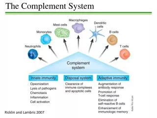
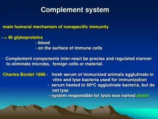
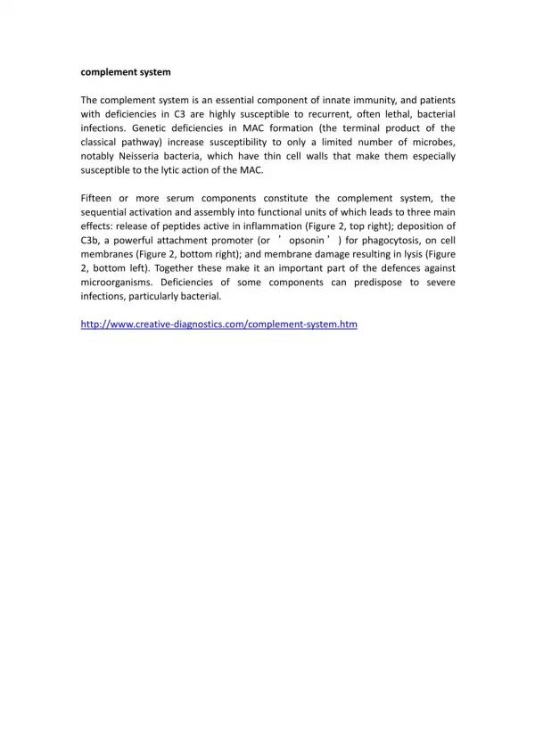
Complement system is a group of heat labile proteins normally found in blood and tissue fluids (except urine and CSF). • They are termed complement as they are required to “complement” the bactericidal effect of antibodies. • They are nine basic factors (C 1 to C 9) which are normally found in an inactive form, activation is through interaction of complement components in a sequential manner (complement activation cascade).
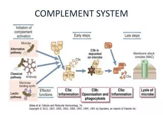
Complement activation pathways Activation of complement factors split them into a small fragment (a) which is considered a by-product and a large (b) factor which continues the activation process. • Classical pathway: Specific (adaptive) immune response • Alternative pathway: Non-specific (innate) immune response • Mannose Binding Lectin pathway: • Similar to alternative pathway in initiation ( being non-specific) • Similar to classical pathway in mechanism of activation.
Classical pathway • Classical pathway is initiated by binding antibody (IgM or IgG [IgG1 and IgG3]) to antigens on microbial cell surface. • C1q (tulip-shape), has 6 binding sites, attaches two or more Fc regions of antibodies. • Binding of C1q activates C1r then C1s (C1 estrase enzyme).
Classical pathway C4b2b3b functions as C5 convertase of the classical pathway
Alternative pathway • C3 is present in plasma, spontaneously hydrolyzed to C3a and C3b but its products are unstable. • Alternative pathway is initiated when C3b is deposited on the pathogen surface (non-specific). • C3bBb, which is stabilized by properdin, functions as C3 convertase of the alternative pathway. • C3bBb3b functions as C5 convertase of the alternative pathway.
Mannose Binding Lectin pathway • Mannose bindinlectin (MBL) pathway is initiated by binding of MBL (a plasma protein) to mannose on pathogen surface (in absence of antibody, non-specific) • MBL is structurally similar to C1q of the classical pathway, it activates MBL-activated serine proteases 1 &2 (MASP1, MASP2) which activate C4. The subsequent steps are similar to the classical pathway.
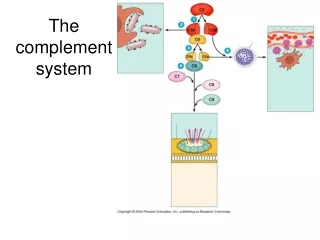
Common pathway Formation of the membrane attack complex (MAC) • C5 convertase of both classical and alternative pathways cleaves C5 into C5a and C5b • C5b attaches to the membrane followed by C6, C7, C8, and C9 forming the membrane attack complex (C5b6789).
Comparison between the three pathways of complement activation
Regulation of the complement system • Complement tends to undergo spontaneous activation, especially by the alternative pathway. These activated complement components can destroy any cell they bind. • It is effective against microbes but not against host cells and tissues because host cells are protected from such damage by regulatory proteins: some are attached to cell surface and some are plasma proteins e.g. C1 inhibitor. • Deficiency of regulatory proteins results in excessive complement activation that causes inflammation and cell death.
Functions of complement • Direct cytolysis: MAC forms a hollow cylinder that is inserted in the host cell membrane allowing passage of water and solutes into the cell leading to cell death (osmotic lysis) e.g. bacterial and tumor cells.
Opsonization: C3b (acts as opsonin) attaches to the antigen, phagocytic cells recognize C3b coated antigen via their C3b receptors (CR1), this facilitates phagocytosis and subsequent intracellular killing.
Functions of complement • Inflammatory response: C5a and C3a have the following biological activities: • Anaphylatoxins: Degranulation of mast cells and basophils to release mediators of inflammation e.g. histamine. • Chemotaxis: recruitment of phagocytic cells to the site of inflammation and stimulation of their phagocytic power and intracellular killing.
Functions of complement • Immune complex clearance: RBCs have C3b receptors, they bind C3b bound to soluble immune complexes and transport them to liver and spleen (rich in fixed phagocytes). These phagocytes, using their own C3b and Fc receptors, remove the immune complexes from RBCs. This helps clearance of soluble immune complexes from the circulation and prevents the development of immune complex diseases. • Activation of B cells: By binding of C3d, generated from C3, to CR2 on B cells